


Foetus Development Illustrations Weeks 1 to 40 - (A PACK OF 40 IMAGES)
Image Description:
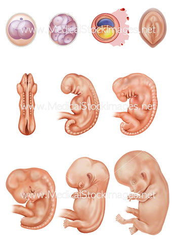
This is the supply of 40 individual illustrations showing the development of the human foetus within the womb. All 40 illustrations can be seen in preview. Once purchased these would be supplied as separate images.
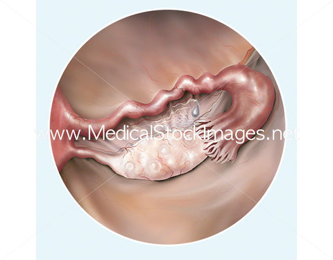
The illustrations start at week 1 and ovulation. Ovulation is the release of an egg from the ovary, into the fallopian tube. Ovulation usually occurs once in every menstrual cycle, when hormone changes trigger an ovary to release an egg.
Week 2 shows cell development where the egg has been fertilised by the male's sperm and the egg divides from a single cell into multiple cells and to a blastocyst ready to implant in the womb. The cell development occurs as the cells travels through the fallopian tube towards the uterus.
Week 3 illustration is split into two parts. The blastocyst has by now implanted into the wall of the uterus and the cells have continued to divide. These two parts show progress in development and the start of differentiation of germ layers. As the third week of development begins, the first signs of the nervous system appear. The two-layered disc of cells becomes a three-layered disc through the process of gastrulation. The embryo takes the shape of an oval-shaped disc and forms an indentation called the primitive streak along the dorsal surface. The second part of the illustration shows further development of the neural plate where the somites and other parts of the mesoderm can be seen through the surface of the ectoderm. The neural plate forms the early brain and spine
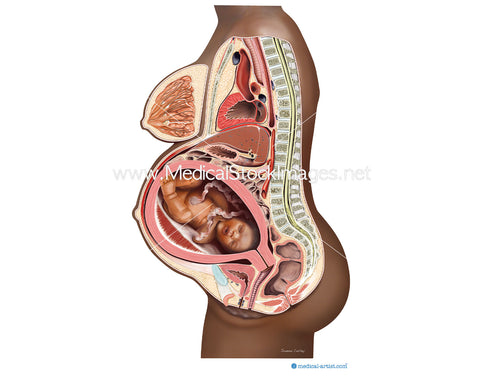
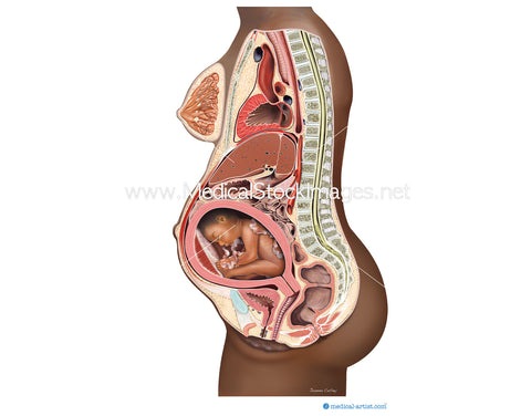
The foetus is shown within the the womb to focus on the foetal development. The embryonic period of the foetal development shows anatomical developments such as arm buds forming into hands, the heart bulge forming into the heart, the development of the organs. The anatomy was researched and each week correctly depicts the amazing developments within the womb.
The developing foetus is shown within the womb to week 20, at week 21 to week 39 the foetus is shown on a black background, at week 40 the baby is born and shown as a newborn.
Image File Sizes:
|
Size |
Pixels |
Inches (@300dpi) |
cm (@300dpi) |
|
Small |
From 600 x 488px |
From 2.0 x 1.5” |
From 5.1 x 3.8cm |
|
Medium |
From 1200 x 1200px |
From 4.0 x 4.0” |
From 10.2 x 10.2cm |
|
Large |
2362 x 2362px |
7.9 x 7.9" |
20.0 x 20.0cm |
Anatomy Visible in the Medical Illustration Includes:
Foetus, fetus, fetal development, womb, baby, conception, uterus, pregnancy, placenta, weeks 1 to 40.
Image created by:
We Also Recommend
