Pencil Drawing Knee Joint Anatomy
Image Description:
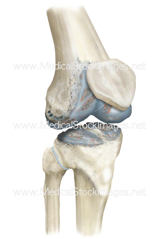
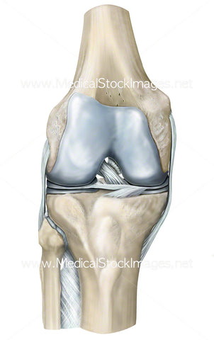
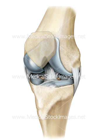
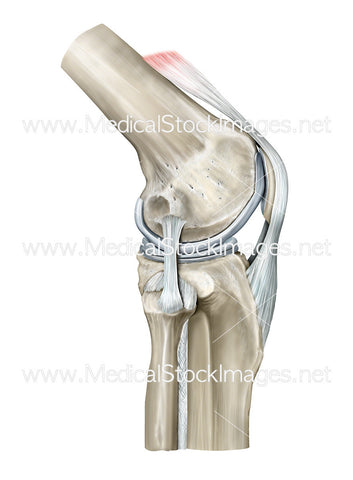
A pencil drawn illustration of the bony anatomy of the knee in anterior knee view. Scanned at a very high resolution this drawing is available to print at up to 45cm in height making it ideal for high impact marketing or artwork for medical practices.
Image File Sizes:
|
Size |
Pixels |
Inches (@300dpi) |
cm (@300dpi) |
|
Small |
400 x 600px |
1.3 x 2.0” |
3.4 x 5.1cm |
|
Medium |
800 x 1200px |
2.7 x 4.0” |
6.8 x 10.2cm |
|
Large |
1600 x 2400px |
5.3 x 8.0” |
13.6 x 20.3cm |
|
X-Large |
2667 x 4000px |
8.9 x 13.3” |
22.6 x 33.9cm |
|
Maximum |
7087 x 10630px |
23.6 x 35.4” |
60.0 x 90.0cm |
Anatomy Visible in the Medical Illustration Includes:
Femur, tibia, fibula, patellofemoral groove, patella groove, patellar sulcus, patellofemoral groove, femoropatellar groove, femoral groove, femoral sulcus, trochlear groove of femur, trochlear sulcus of femur, trochlear surface of femur, or trochlea of femur, medical epicondyle, medical collateral ligament, posterior cruciate ligament, medical meniscus, transverse ligament, interosseous, ligament of fibula head, tibial plateau, lateral meniscus, anterior cruciate ligament, lateral cruciate ligament, articular cartilage.
Image created by:
We Also Recommend