Cervical Spine in Anterior View
Image Description:
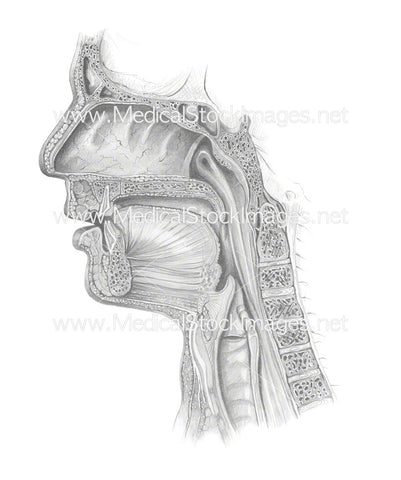
Illustration of the cervical spine in anterior view. The cervical spine contains 7 vertebrae, named from C1 to C7 descending from the top of the neck to bottom. The cervical spine is more flexible and mobile than the thoracic or lumbar regions of the spine. However they are also comprised of thinner and more delicate bones.
C1 and C2 are commonly known as the atlas and axis. The atlas forms the joint connecting the skull and spine. The lowest cervical vertebra is C7, or vertebra prominens, has a long and prominent spinous process.
Image File Sizes:
|
Size |
Pixels |
Inches |
cm |
|
Small |
364x600px |
1.2x2.0” @300dpi |
3.1x5.1cm @300dpi |
|
Medium |
727x1200px |
2.4x4.0” @300dpi |
6.2x10.2cm @300dpi |
|
Large |
1793x2960px |
6.0x9.9” @300dpi |
15.2x25.1cm @300dpi |
Anatomy Visible in the Medical Illustration Includes:
Cervical vertebra, neck, vertebral body, atlas, axis, C1, C2, C3, C4, C5, C6, C7, vertebra prominens, spinous process, sulcus for spinal nerve, vertebral body, anterior tubercle, posterior tubercle, transverse process, uncinate process, inferior articular process, superior articular process.
Image created by:
We Also Recommend