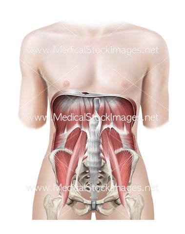
Arteries and Veins of the Thoracic and Abdominal Region
Image Description:
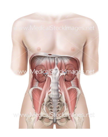
Anterior view of the ventral cavity of muscle anatomy with the descending aorta. The descending aorta originates in the aortic arch of the heart. There are two sections to the decending aorta – the thoracic and abdominal aorta.
Image File Sizes:
|
Size |
Pixels |
Inches |
cm |
|
Small |
476x600px |
1.6x2” @300dpi |
4x5.1cm @300dpi |
|
Medium |
951x1200px |
3.2x4” @300dpi |
8.1x10.2cm @300dpi |
|
Large |
1901x2400px |
6.3x8” @300dpi |
16.1x20.3cm @300dpi |
|
X-Large |
3168x4000px |
10.6x13.3” @300dpi |
26.8x33.9cm @300dpi |
|
Maximum |
6916x8731px |
23.1x29.1” @300dpi |
58.6x73.9cm @300dpi |
Anatomy Visible in the Medical Illustration Includes:
Quadratus lumborum muscle, transverse abdominis muscle, psaos major muscle, greater trochanter of femur, iliopsoas muscle, pelvis, intercostal muscles, subcotal muscles, external intercostal muscles, costal part of diaphragm, kidneys, tensor fasciae latae, Sartorius, descending aorta, inferior vena cava, common iliac artery, common iliac vein.
Image created by:
We Also Recommend