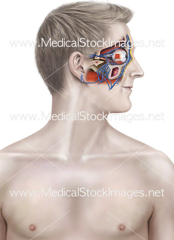
Anatomy of the Eye and Eyelid in Cross-section
Image Description:
Illustration of the eye and upper and lower eyelids in cross-section.
Image File Sizes:
|
Size |
Pixels |
Inches (@300dpi) |
cm (@300dpi) |
|
Small |
600 x 564px |
2.0 x 1.9” |
5.1 x 4.8cm |
|
Medium |
1200 x 1127px |
4.0 x 3.8” |
10.2 x 9.5cm |
|
Large |
3074 x 2886px |
10.2 x 9.6” |
26.0 x 24.4cm |
Anatomy Visible in the Medical Illustration Includes:
Iris, lens, cornea, anterior chamber, chamber angle, corneoscleral limbus, ciliary body, ciliary muscle, zonular fibers, ora serrata, vitreous body, lateral rectus, retina, choroid, sclera, fovea centralis, optic nerve, lamina cribosa, optic disk, medial rectus, hyaloids, fossa, ocular conjuctiva, pigment epithelium, canal of schlemm, posterior chamber, upper eyelid, lower eyelid, orbicularis oculi, orbital septum, superior tarsal muscle, inferior tarsal muscle, orbital roof, periorbita.
Image created by:
We Also Recommend