Anatomy of the Large Bowel with Intestinal Arteries
Image Description:
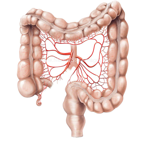
Anatomy of the large bowel with intestinal arteries. The abdominal aorta is the primary blood supply to the intestines from three pairs of unpaired branches. These are the celiac artery and the superior and inferior mesenteric arteries supplying blood to the left third and the transverse colon and to the sigmoid colon.
Image File Sizes:
|
Size |
Pixels |
Inches (@300dpi) |
cm (@300dpi) |
|
Small |
600 x 600px |
1.5 x 1.5” |
3.8 x 3.8cm |
|
Medium |
1200 x 1200px |
3.0 x 3.0” |
7.6 x 7.6cm |
|
Large |
2400 x 2400px |
6.0 x 6.0” |
15.2 x 15.2cm |
|
X-Large |
4000 x 4000px |
10.0 x 10.0” |
25.4 x 25.4cm |
|
Maximum |
6772 x 6772px |
16.9 x 16.9” |
43.0 x 43.0cm |
Anatomy Visible in the Medical Illustration Includes:
Transverse colon, ascending colon, descending colon, aorta, superior mesenteric artery, inferior mesenteric artery, sigmoid arteries, right colic artery, middle colic artery, rectal artery.
Image created by:
We Also Recommend


