Hip
A healthy hip carries almost 5 times a person’s body weight during an everyday activity such as walking. These medical illustrations of the hip include healthy anatomy and surgical repairs to the hip. Hip surgery illustrations provided include hip replacement and resurfacing.
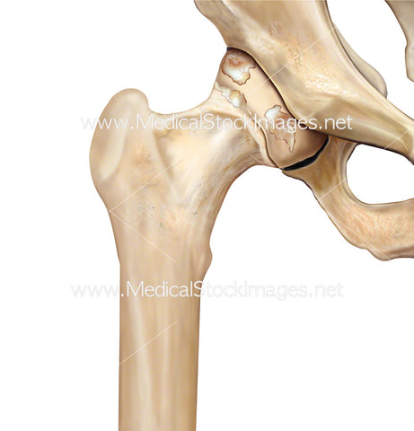
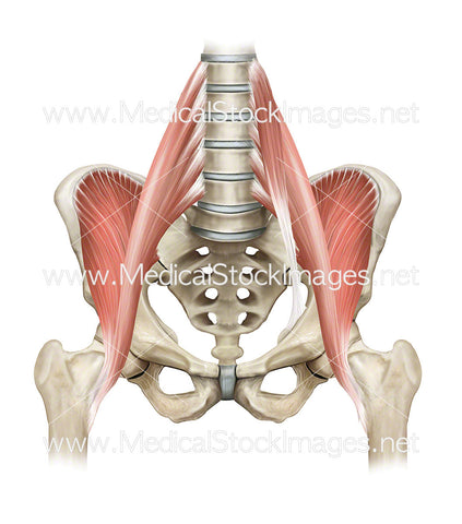
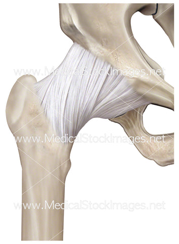
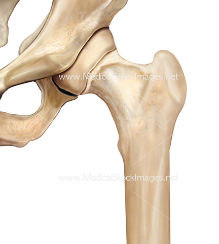
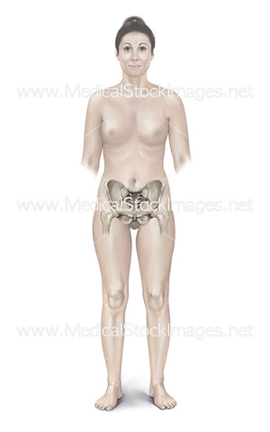
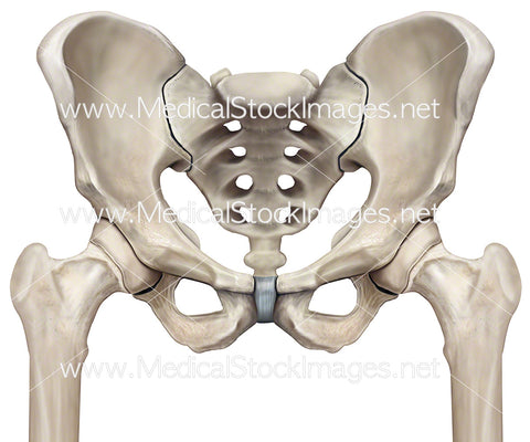
Hip Anatomy
There are five different muscle groups that cover or are attached to the hip joint and generate different kinds of movements, such as:
- Gluteals: The ones in your buttocks, which are subdivided into three different parts: maximus, minimus and medius.
- Quadriceps: These are four different muscles, (vastus lateralis, medialis, intermedius and rectus femoris), located at the front of the femur.
- Iliopsoas: The primary hip flexor muscle that connects the lower part of the spine with the pelvis.
- Hamstrings: Three muscles at the back of the thigh.
- Groin: Also known as the adductor muscle, which are attached to the pubis.
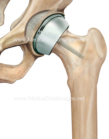
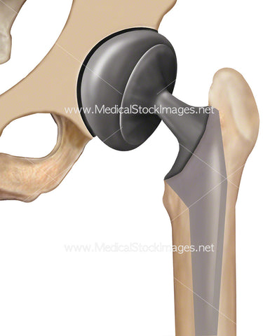
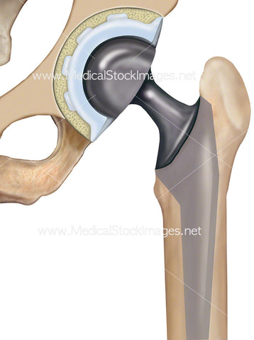
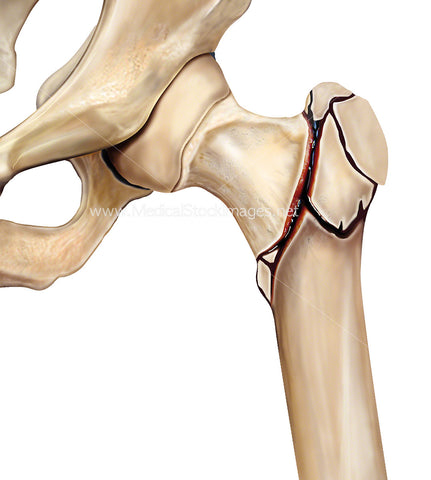
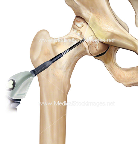
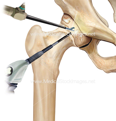
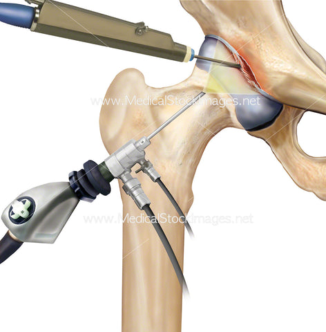
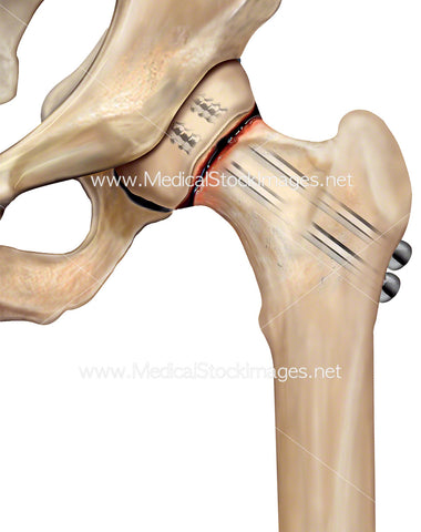
Hip Illustrations Include:
Hip fractures, partial hip replacement, full hip replacement, articular capsule, arthroscopy, subcapital hip fracture and its repair and hip interochanteric fracture repair.