Anatomy of Inner and Outer Ear
Image Description:
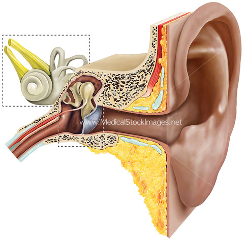
Anatomy of inner and outer ear shown within their relevant areas by use of call out boxes.
Image File Sizes:
|
Size |
Pixels |
Inches (@300dpi) |
cm (@300dpi) |
|
Small |
600 x 592px |
2.0 x 2.0” |
5.1 x 5.0cm |
|
Medium |
1200 x 1183px |
4.0 x 3.9” |
10.2 x 10.0cm |
|
Large |
2131 x 2100px |
7.1 x 7.0” |
18.0 x 17.8cm |
Anatomy Visible in the Medical Illustration Includes:
External ear, auricle, ear canal, outer ear, middle ear, inner ear, ear drum, malleus, semi-circular canals, incus, stapes, vestibular nerve, facial nerve, cochlea, eustachian tube.
Image created by:
We Also Recommend


