Anatomy of Healthy Skin at a Cellular Level
Image Description:
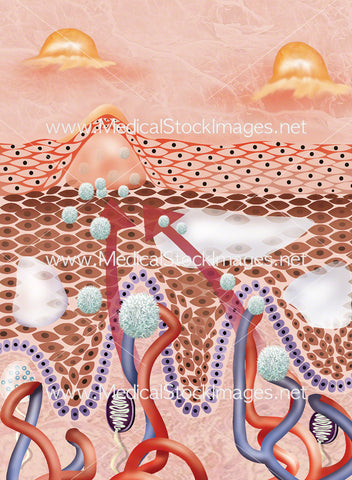
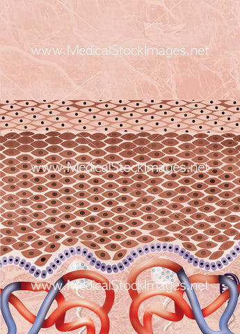
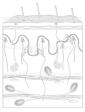
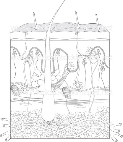
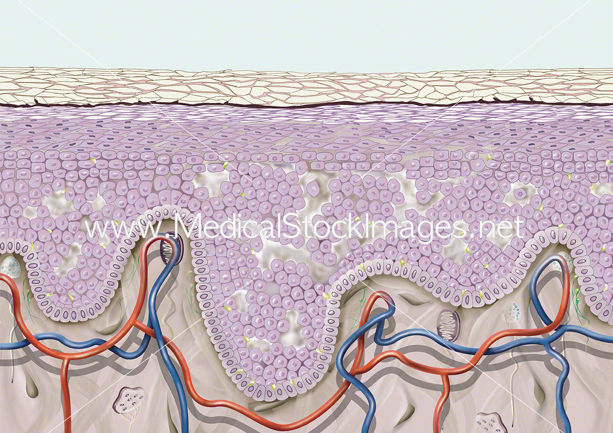
Cross section of the skin showing healthy or young skin at a cellular level. There are three layers of skin – the epidermis (the outer layer), the dermis (under the epidermis) and the subcutaneous tissue layer (hypodermis). The skin is the largest organ of the human body.
Image File Sizes:
|
Size |
Pixels |
Inches |
cm |
|
Small |
600x425px |
2.0x 1.4” @300dpi |
5.1x3.6cm @300dpi |
|
Medium |
1200x849px |
4.0x2.8” @300dpi |
10.2x7.2cm @300dpi |
|
Large |
2400x1697px |
8.0x5.7” @300dpi |
20.3x14.4cm @300dpi |
|
X-Large |
4000x2828px |
13.3x9.4” @300dpi |
33.9x23.9cm @300dpi |
|
Maximum |
4961x3508px |
16.5x11.7” @300dpi |
42x29.7cm @300dpi |
Anatomy Visible in the Medical Illustration Includes:
Skin surface, epidermis, dermis, subcutaneous tissue, wrinkle, aged skin, epidermal atrophy, reduced and disorganised collagen fibers, reduced vascular tissue, elastic fibers, free nerve endings.
Image created by:
We Also Recommend