Fertilization (Fertilisation) and Cell Development
Image Description:
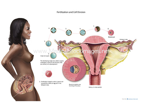
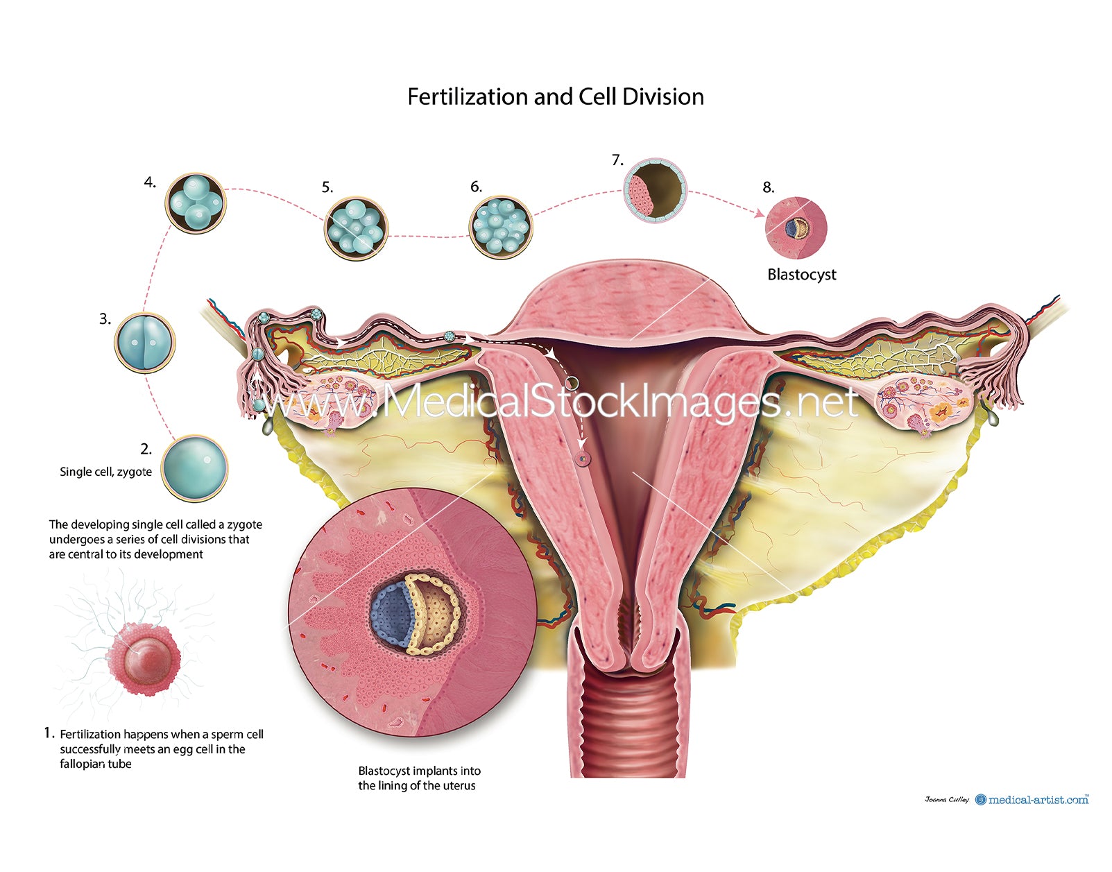
The illustration depicts fertilization (fertilisation) and cell development. The purpose of the illustration to show the sperm fertilizing an egg and subsequent cell development to blastocyst stage. Fertilization occurs usually in the fallopian tube and by the time the blastocyst has developed it has reached the internal structure of the uterus (womb) and implants into the layers of the uterus ready for the next stage of foetal (fetal) development.
Image File Sizes:
|
Size |
Pixels |
Inches (@300dpi) |
cm (@300dpi) |
|
Small |
885px x 600px |
3" x 2” |
7cm x 5cm |
|
Medium |
1770px x 1200px |
6" x 4” |
15 x 10cm |
|
Large |
2400 x 1627 px |
8" x 5” |
20cm x 14cm |
|
X-Large |
4000px x 2712px |
13" x 9” |
34cm x 23cm |
|
Maximum |
5899px x 4000 px |
20" x 13” |
50cm x 39 cm |
Anatomy Visible in the Medical Illustration Includes:
Lungs, diaphragm, ribs, intercostal , muscles, bronchus, bronchii, bronchioles, aortic hiatis, transverse muscles, illiacus, sacrum, pelvis, rectus femoris, tensor fascia latae, psoas, esophageal hiatus aorta, heart.
Image created by:
We Also Recommend