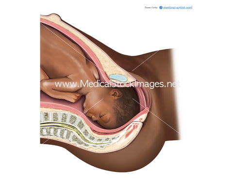
Internal Foetus Monitoring during Pregnancy (African Heritage).
Image Description:
The internal anatomy has been illustrated within the body of a female and her baby of African heritage. The process of internal fetal heart rate monitoring uses an electronic transducer connected directly to the fetal skin. A wire electrode is attached to the fetal scalp or other body part through the cervical opening and is connected to the monitor. This type of electrode is sometimes called a spiral or scalp electrode.
Image File Sizes:
|
Size |
Pixels |
Inches (@300dpi) |
cm (@300dpi) |
|
Small |
600px x 609px |
2" x 2” |
5cm x 5cm |
|
Medium |
1183px x 1200px |
4 x 4” |
10 x 10cm |
|
Large |
2366 x 2400 px |
8" x 8” |
20cm x 20 cm |
|
X-Large |
3943px x 4000 px |
13" x 13” |
33cm x 33cm |
|
Maximum |
4926px x 5000 px |
16" x 16” |
41cm x 41 cm |
Anatomy Visible in the Medical Illustration Includes:
Lungs, diaphragm, ribs, intercostal , muscles, bronchus, bronchii, bronchioles, aortic hiatis, transverse muscles, illiacus, sacrum, pelvis, rectus femoris, tensor fascia latae, psoas, esophageal hiatus aorta, heart.
Image created by:
We Also Recommend


