


Foetus Development Weeks 1 to 40 Including Female Body Labelled (Multipack of 40 IMAGES).
Image Description:
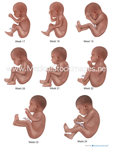
Development of the human foetus during pregnancy shown in context to the mother. The illustrations have additional labelling.
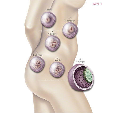
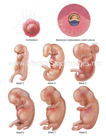
Week 1 shows cell development and the fertilised cell as it divides from a single cell into multiple cells and to a blastocyst ready to implant in the womb.
Week 2 is the blastocyst implanting into the wall of the uterus (womb). When implantation succeeds the blastocyst adheres to the endometrium part of the womb.
Week 3 illustration is split into two parts. These two parts show the differentiation of germ layers. As the third week of development begins, the first signs of the nervous system appear. The two-layered disc of cells becomes a three-layered disc through the process of gastrulation. The embryo takes the shape of an oval-shaped disc and forms an indentation called the primitive streak along the dorsal surface. The second part of the illustration shows further development of the neural plate where the somites and other parts of the mesoderm can be seen through the surface of the ectoderm. The neural plate forms the early brain and spine.
Image File Sizes:
|
Size |
Pixels |
Inches (@300dpi) |
cm (@300dpi) |
|
Small |
600 x 600px |
2.0 x 2.0” |
5.1 x 5.1cm |
|
Medium |
1200 x 1200px |
4.0 x 4.0” |
10.2 x 10.2cm |
|
Large |
2400 x 2400px |
8.0 x 8.0” |
20.3 x 20.3cm |
|
X-Large |
2942 x 2942px |
9.8 x 9.8” |
24.9 x 24.9cm |
Anatomy Visible in the Medical Illustration Includes:
Foetus, fetus, fetal development, womb, cells, blastocyst, baby, embryo, neural tube, conception, uterus, fertilized egg, 2-cell stage, 4-cell stage, 8-cell stage, 16 cell-stage, neural plate, neural groove.
Image created by:
We Also Recommend