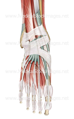
Muscles and Tendons of the Foot in Dorsal View
Image Description:
Illustration of the superior muscles and tendon sheaths of the foot in dorsal view.
Image File Sizes:
|
Size |
Pixels |
Inches |
cm |
|
Small |
250x600px |
0.8x2.0” @300dpi |
2.1x5.1cm @300dpi |
|
Medium |
499x1200px |
1.7x4.0” @300dpi |
4.2x10.2cm @300dpi |
|
Large |
998x2400px |
3.3x8.0” @300dpi |
8.5x20.3cm @300dpi |
|
X-Large |
1731x4164px |
5.8x13.9” @300dpi |
14.7x35.3cm @300dpi |
Anatomy Visible in the Medical Illustration Includes:
Muscular foot, fiborous tendon sheaths, triceps surae, tibialis anterior, tibia, extensor hallucis longus, medial malleolus, inferior extensor retinaculum, extensor hallucis brevis, extensor digitorum brevis, extensor digitorum longus, interossei, extensor hallucis longus, abductor digiti minimi, tuberosity of fifth metatarsal, fibularis tertius, fibularis brevis, lateral malleolus, superior extensor retinaculum, fibularis brevis, extensor digitorum longus, fibularis longus.
Image created by:
We Also Recommend


