Heart Sternocostal Surface
Image Description:
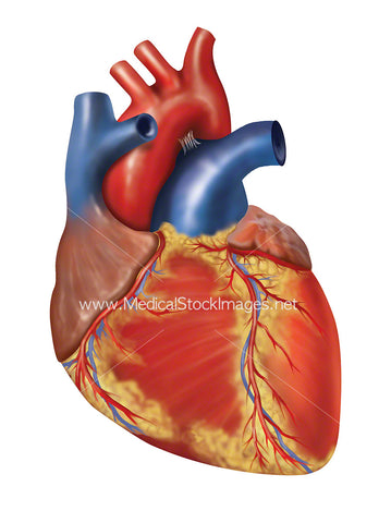
Sternocostal or anterior surface anatomy of a healthy heart. It consists of the anterior surfaces of the right atrium and right ventricle, and small parts of the left atrium and left ventricle.
Image File Sizes:
|
Size |
Pixels |
Inches |
cm |
|
Small |
428x600px |
1.4x2.0” @300dpi |
3.6x5.1cm @300dpi |
|
Medium |
855x1200px |
2.9x4.0” @300dpi |
7.2x10.2cm @300dpi |
|
Large |
1711x2400px |
5.7x8.0” @300dpi |
14.5x20.3cm @300dpi |
|
X-Large |
2851x4000px |
9.5x13.3” @300dpi |
24.1x33.9cm @300dpi |
|
Maximum |
3750x5261px |
12.5x17.5” @300dpi |
31.8x44.5cm @300dpi |
Anatomy Visible in the Medical Illustration Includes:
Heart surface, superior vena cava, ascending aorta, arch of aorta, right atrium, right ventricle, left ventricle, left atrium, pulmonary veins, pulmonary trunk, left auricle, inferior interventricular branch or left coronary artery, great cardiac vein, apex, anterior interventricular groove, inferior margin.
Image created by:
We Also Recommend