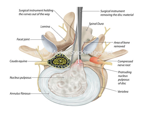
A Diagram Showing a Disc Bulge Being Removed-Labelled
Image Description:
Illustration of a lumbar vertebra in anterolateral view with a compressed nerve trapped by a bulging or herniated disc. A bulging lumbar disc occurs when the soft inner material of a spinal disc in the lower back bulges out through a tear in the outer layer, trapping a nerve and causing pain. Common symptoms include low back pain, and pain, numbness, tingling, or weakness that radiates down the leg (sciatica). Surgery is performed to remove the protruding disc material and to relieve the compression on the trapped nerve.
Image File Sizes:
|
Size |
Pixels |
Inches |
cm |
|
Small |
600x516px |
2.0x1.7” @300dpi |
5.1x4.47cm @300dpi |
|
Medium |
1395x1200px |
4.65x4” @300dpi |
11.8x10.16cm @300dpi |
|
Large |
3038x2613px |
10.13"X8.71” @300dpi |
25.72x22.12cm @300dpi |
Anatomy Visible in the Medical Illustration Includes:
Lumbar vertebra, vertebral body, pedicle, transverse process, superior articular process, vertebral foramen, accessory process, spinous process, bulging disc, compressed nerve, disc, disc protrusion, disc bulge, herniated disc, sciatica, nerve pain, facet joint.
Image created by:
We Also Recommend

