Anatomy of a Common Wart Illustration
Image Description:
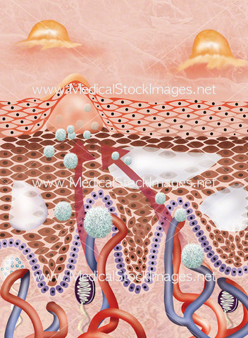
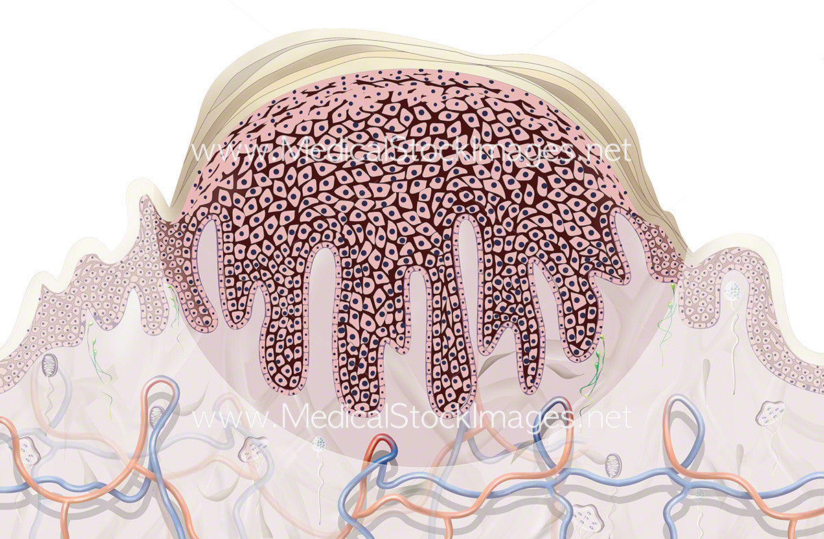
Depiction of a common wart verruca vulgaris or palmer wart or junior wart. Illustrated in cross section at a highly magnified view at cellular level to show the characteristic appearance as if under the microscope. Shown to have a thickening of the stratum corneum known as hyperkeratosis which is the abnormal thickening of the outer layer of the skin. A thickening of the stratum spinosum known as epidermal hyperplasia. A thickening of the stratum granulosum as well as rete ridge elongation and large blood vessels at the dermoepidermal junction which is the area of tissue that joins the epidermal and dermal layers of the skin. Warts are usually caused by an infection of the top layer of skin by viruses from the human papillomavirus or HPV family.
Image File Sizes:
|
Size |
Pixels |
Inches (@300dpi) |
cm (@300dpi) |
|
Small |
600 x 393px |
2 x 1.3” |
5.1 x 3.3cm |
|
Medium |
1200 x 785px |
4 x 2.6” |
10.2 x 6.7cm |
|
Large |
2400 x 1570px |
8 x 5.2” |
20.3 x 13.3cm |
|
X-Large |
2914 x 1906px |
9.7 x 6.4” |
24.7 x 16.1cm |
Anatomy Visible in the Medical Illustration Includes:
Stratum corneum, stratum spinosum, stratum granulosum, epidermis, dermoepidermal junction, large blood vessels, rete ridge, verruca vulgaris, common wart, junior wart.
Image created by:
We Also Recommend