Posterior View of the Spinal Cord and the Meninges
Image Description:
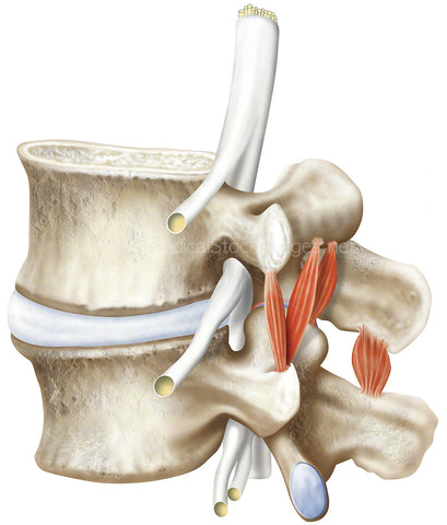
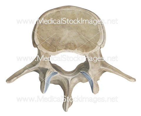
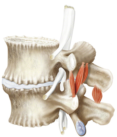
The illustration depicts the posterior view of the pelvis with the spinal cord within the lumbar region. Cross sectioned to reveal the three layers of protective tissue membrane surrounding the cord called the meninges. These are the dura mater, arachnoid mater and pia mater.
Image File Sizes:
|
Size |
Pixels |
Inches |
cm |
|
Small |
418x600px |
1.4x2.0” @300dpi |
3.5x5.1cm @300dpi |
|
Medium |
835x1200px |
2.8x4.0” @300dpi |
7.1x10.2cm @300dpi |
|
Large |
1670x2400px |
5.6x8.0” @300dpi |
14.1x20.3cm @300dpi |
|
X-Large |
2783x4000px |
9.3x13.3” @300dpi |
23.6x33.9cm @300dpi |
|
Maximum |
3870x5433px |
12.6x18.1” @300dpi |
32.0x46.0cm @300dpi |
Anatomy Visible in the Medical Illustration Includes:
Lumbar vertebra, L4, epidural space, arachnoid mater, anterior vertebral venous plexus, dura mater, conus medullaris, posterior vertebral venous plexus, meningeal layers, spinal cord, anterior spinal artery, anterior spinal vein, pia mater, denticulate ligament, arachnoid mater, subdural space, cauda equina, spinal arachnoid mater, subarachnoid space.
Image created by:
We Also Recommend