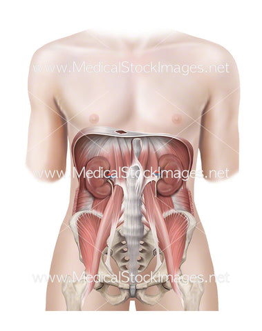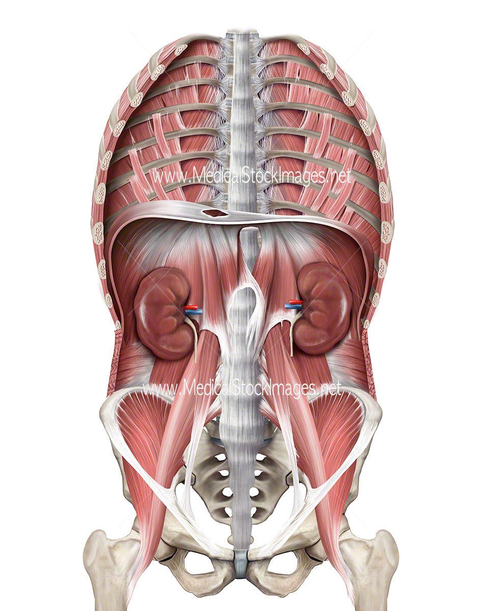
Illustration of Thoracic Region Including Diaphragm, Pelvis, Kidneys
Image Description:
Anterior view of the ventral cavity of muscle anatomy from the pelvic region to the rib cage including the positioning of the kidneys.
Image File Sizes:
|
Size |
Pixels |
Inches (@300dpi) |
cm (@300dpi) |
|
Small |
476 x 600px |
1.6 x 2.0” |
4.0 x 5.1cm |
|
Medium |
951 x 1200px |
3.2 x 4.0” |
8.1 x 10.2cm |
|
Large |
1901 x 2400px |
6.3 x 8.0” |
16.1 x 20.3cm |
|
X-Large |
3168 x 4000px |
10.6 x 13.3” |
26.8 x 33.9cm |
|
Maximum |
6916 x 8731px |
23.1 x 29.1” |
58.6 x 73.9cm |
Anatomy Visible in the Medical Illustration Includes:
Pelvis, psaos major muscle, psaos minor muscle iliacus muscle, greater trochanter of femur, pelvis, iliopsoas muscle, esophageal aperture, transverse abdominis, left crus, median arcuate ligament, lateral arcuate ligament, medial arcuate ligament, vena cava aperture, central tendon, intercostal muscles, subcostal muscles, external intercostal muscles, costal part of diaphragm, diaphragm, kidneys, trunk wall, muscles of trunk wall.
Image created by:
We Also Recommend