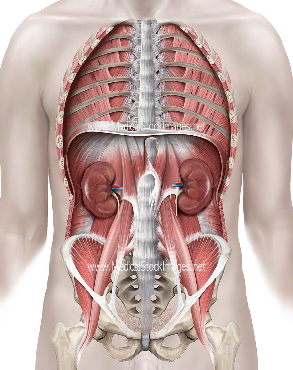
Kidney Positioning within Ventral Cavity
Image Description:
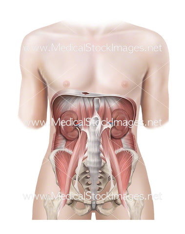
Anterior view of the ventral cavity. Anatomy removed to show just the kidneys and their position within the human body.
Image File Sizes:
|
Size |
Pixels |
Inches (@300dpi) |
cm (@300dpi) |
|
Small |
476 x 600px |
1.6 x 2.0” |
4.0 x 5.1cm |
|
Medium |
951 x 1200px |
3.2 x 4.0” |
8.1 x 10.2cm |
|
Large |
1901 x 2400px |
6.3 x 8.0” |
16.1 x 20.3cm |
|
X-Large |
3168 x 4000px |
10.6 x 13.3” |
26.8 x 33.9cm |
|
Maximum |
6916 x 8731px |
23.1 x 29.1” |
58.6 x 73.9cm |
Anatomy Visible in the Medical Illustration Includes:
Quadratus lumborum muscle, transverse abdominis muscle, psaos major muscle, greater trochanter of femur, iliopsoas muscle, pelvis, intercostal muscles, subcostal muscles, external intercostal muscles, costal part of diaphragm, kidneys.
Image created by:
We Also Recommend