Anatomy of the Brainstem
Image Description:
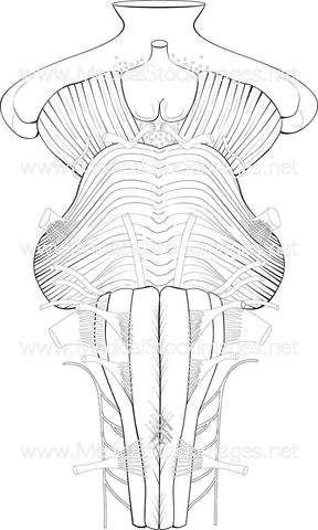
Illustrations showing ventral (anterior) view of the brainstem. The brainstem is divided into three section – the midbrain, the pons and the medullar.
Image File Sizes:
|
Size |
Pixels |
Inches (@300dpi) |
cm (@300dpi) |
|
Small |
360 x 600px |
1.2 x 2.0” |
3.1 x 5.1cm |
|
Medium |
720 x 1200px |
2.4 x 4.0” |
6.1 x 10.2cm |
|
Large |
1439 x 2400px |
4.8 x 8.0” |
12.2 x 20.3cm |
|
X-Large |
2026 x 3379px |
6.8 x 11.3” |
17.2 x 28.6cm |
Anatomy Visible in the Medical Illustration Includes:
Brainstem, brain, midbrain, medullar, pons, ventral, anterior, optic chiasm, infundibulum, tuber cinereum, mammillary body, oculomotor nerve, trochlear nerve, trigeminal nerve, abducens nerve, facial nerve, vestibulocochlear nerve, glossopharyngeal nerve, vagus nerve, decussation of pyramids, circumolivary bundle, pyramid, olive, hypoglossal nerve, pons, middle cerebellar peduncle, lateral geniculate nucleus, medial geniculate nucleus, optic nerve
Image created by:
We Also Recommend

