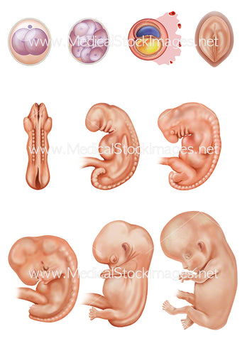
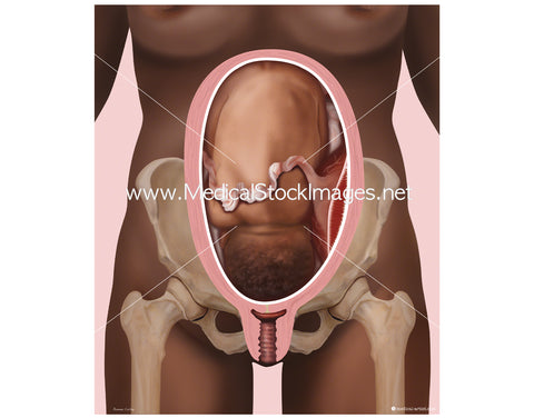
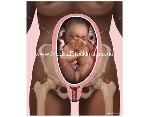
Embryonic Development from Cell Development to Week 8 (part of a set with a white baby)
Image Description:
The developing embryo from fertilisation to week 8. Showing the embryogenesis process which is the process by which the embryo forms and develops. The illustrations created by a medical illustrator with emphasis on the depiction of clinical accuracy. Each stage researched and these illustrations show the fetal (foetal) development from the date of conception. Alternatively, a clinician calculates a pregnancy of a mothers estimated due date, counting ahead 40 weeks from the start of their patients last period. This works out usually as a pregnancy dated two weeks ahead of actual conception.
In this order from the top and left to right, fertilization, (fertilisation), implantation of a blastocyst within the womb lining of the womb, week 3, week 4, week 5, week 6, week 7, week 8. Image File Sizes:
|
Size |
Pixels @300dpi |
Inches (@300dpi) |
cm (@300dpi) |
|
Small |
435px x 600px @300dpi |
1.4" x 2.0” @300dpi |
4cm x 5.1cm @300dpi |
|
Medium |
869px x 1200px @300dpi |
3" x 4.0” @300dpi |
7cm x 10cm @300dpi |
|
Large |
1738px x 2400px @300dpi |
5.8" x 8.0” @300dpi |
14.7cm x 20.3cm @300dpi |
|
X-Large |
2480px x 3425px @300dpi |
8.3" x 11.4” @300dpi |
21.0cm x 29.0cm @300dpi |
Anatomy Visible in the Medical Illustration Includes:
Embryogenesis, embryo, cell, cells, division, zygote, cleavage, blastocyst, bilaminar disc formation, disc, implantation, week 3, week 4, week 5, week 6, week 7, week 8, foetus, fetus, fetal, baby.
Image created by:
We Also Recommend

