Fertilisation and Cell Development Featuring a woman of African Heritage.
Image Description:
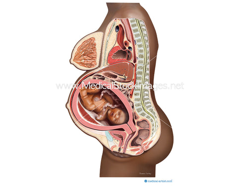
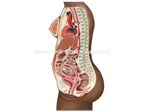
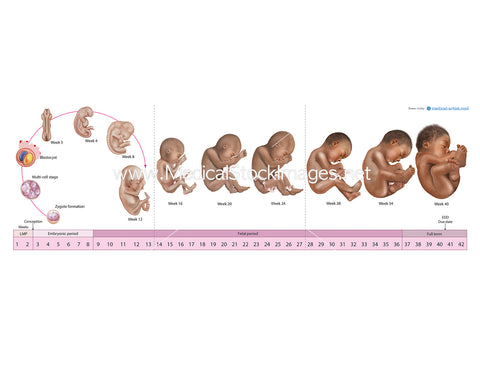
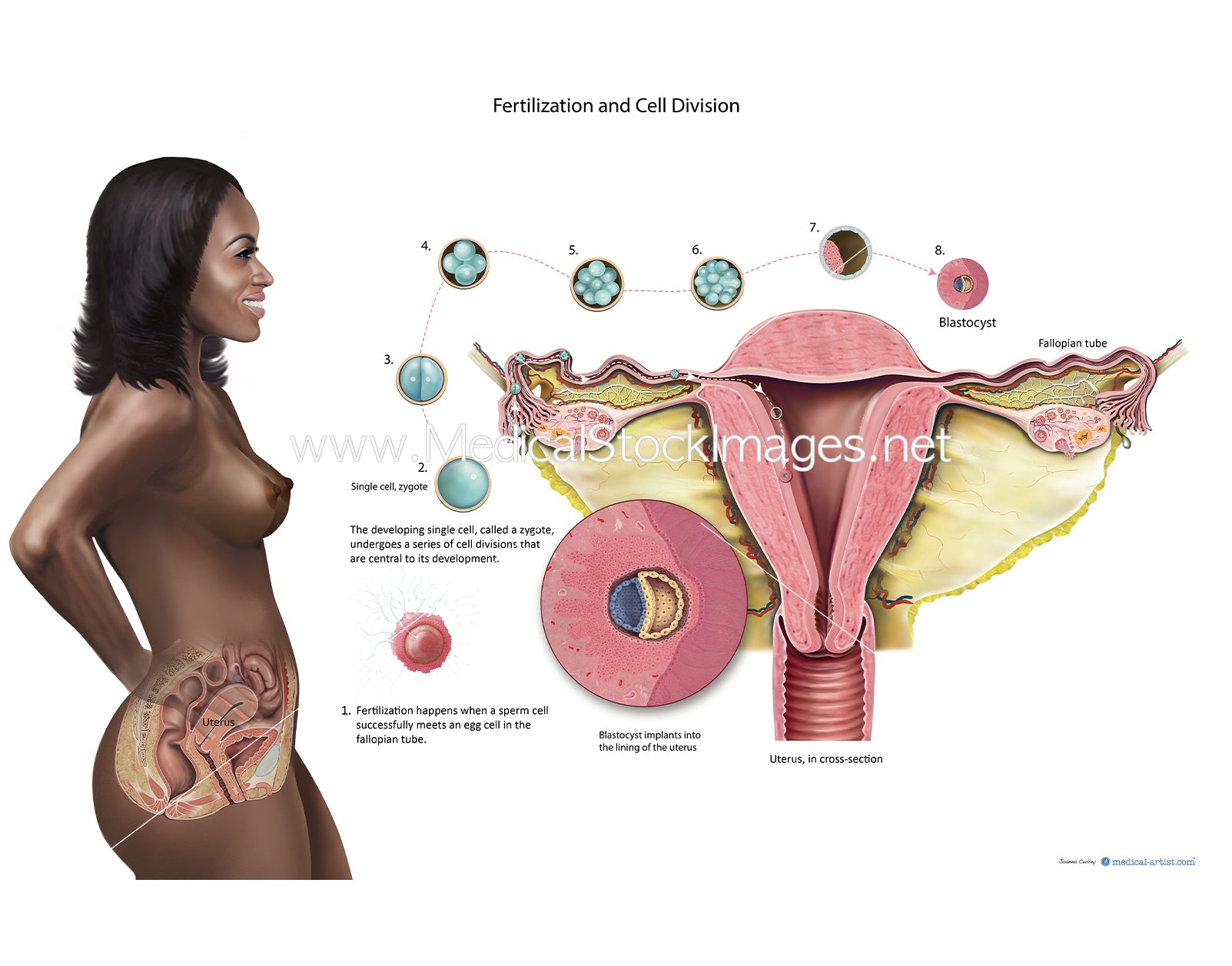
The illustration depicts fertilisation (fertilization) and cell development with the reproductive anatomy visible on a background female of African heritage. The purpose of the illustration to show the the sperm fertilising an egg and subsequent cell development to blastocyst stage. Fertilisation occurs usually in the fallopian tube and by the time the blastocyst has developed it has reached the internal structure of the uterus *womb and implants into the layers of the uterus wall or lining.
Image File Sizes:
|
Size |
Pixels |
Inches (@300dpi) |
cm (@300dpi) |
|
Small |
885px x 600px |
3" x 2” |
7cm x 5cm |
|
Medium |
1770px x 1200px |
6" x 4” |
15 x 10cm |
|
Large |
2400 x 1627 px |
8" x 5” |
20cm x 14cm |
|
X-Large |
4000px x 2712px |
13" x 9” |
34cm x 23cm |
|
Maximum |
5899px x 4000 px |
20" x 13” |
50cm x 39 cm |
Anatomy Visible in the Medical Illustration Includes:
Lungs, diaphragm, ribs, intercostal , muscles, bronchus, bronchii, bronchioles, aortic hiatis, transverse muscles, illiacus, sacrum, pelvis, rectus femoris, tensor fascia latae, psoas, esophageal hiatus aorta, heart.
Image created by:
We Also Recommend