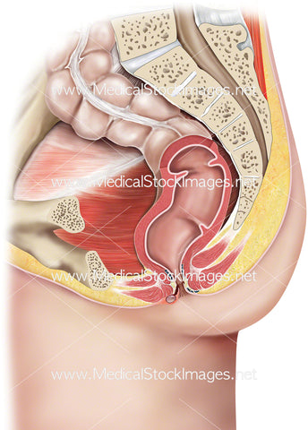
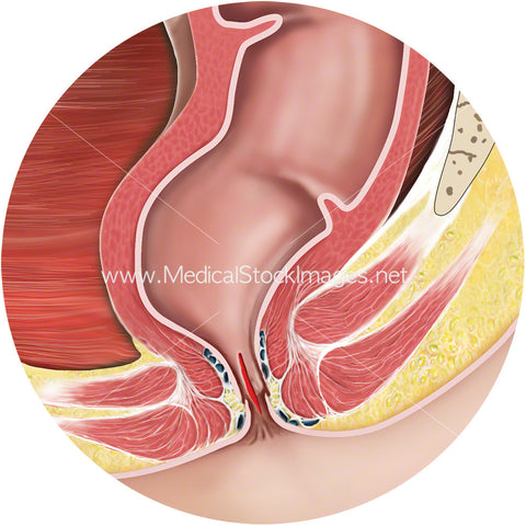
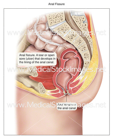
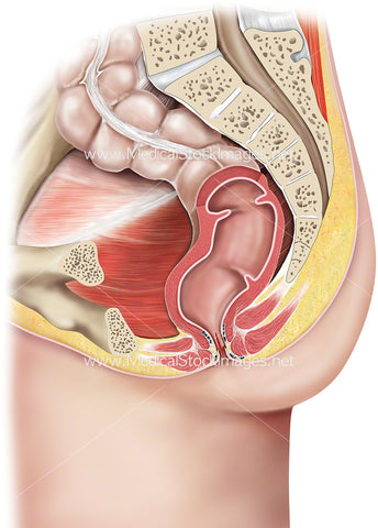
Large Bowel Showing Cross Section of Caecum
Image Description:
The large bowel has been illustrated to show the four sections that is the ascending colon, the transverse colon, the descending colon and the sigmoid colon. The caecum has been opened in cross section to reveal where the large bowel joins the end of the small intestine at the cecum, via the ileocecal valve. The blood supply and the mesentry (in yellow) have also been included. The mesentery is the folds of the peritoneum encloses the intestines and attaches them to the posterior abdominal wall.
Image File Sizes:
|
Size |
Pixels |
Inches (@300dpi) |
cm (@300dpi) |
|
Small |
600 x 600px |
2.0 x 2.0” |
5.1 x 5.1cm |
|
Medium |
1200 x 1200px |
4.0 x 4.0” |
10.2 x 10.2cm |
|
Large |
2400 x 2400px |
8.0 x 8.0” |
20.3 x 20.3cm |
|
X-Large |
4000 x 4000px |
13.3 x 13.3” |
33.9 x 33.9cm |
|
Maximum |
6772 x 6772px |
22.6 x 22.6” |
57.3 x 57.3cm |
Anatomy Visible in the Medical Illustration Includes:
Large, bowel, caecum, colon, ascending, transverse, descending, sigmoid, intestine, ileocecal, valve, mesentery.
Colon, cancer, bowel cancer, rectal cancer, middle colic artery, superior mesenteric artery, inferior mesenteric artery, descending colon, right hemi colectomy, anastomosis, right extended colectomy, splenic flexure, Crohn’s disease.
Image created by:
We Also Recommend


