Anatomy of the Spinal Cord & its Meningeal Layers (Labelled in English)
Image Description:
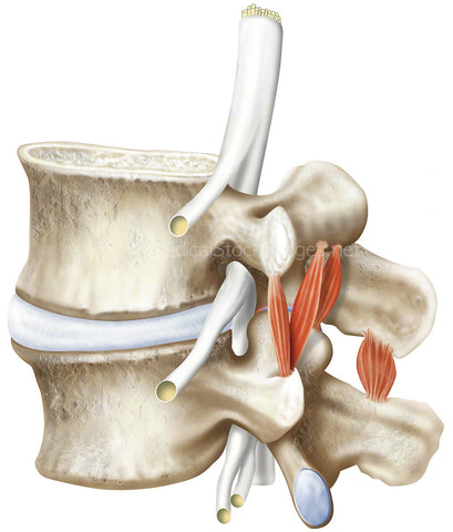
The illustration depicts the three layers of protective tissue membrane called the meninges which are called the dura mater, arachnoid mater, and pia mater. They surround the neuraxis and line the cranium and vertebral canal. Along side this is single vertebra lumbar 4 (L4) to gain a superior view down onto the spinal cord.
Image File Sizes:
|
Size |
Pixels |
Inches |
cm |
|
Small |
472x600px |
1.6x2” @300dpi |
4x5.1cm @300dpi |
|
Medium |
944x1200px |
3.1x 4” @300dpi |
8x10.2cm @300dpi |
|
Large |
1888x2400px |
6.3x8” @300dpi |
16x20.3cm @300dpi |
|
X-Large |
2759x3508px |
9.2x11.7” @300dpi |
23.4x29.7cm @300dpi |
Anatomy Visible in the Medical Illustration Includes:
Lumbar vertebra, L4, epidural space, arachnoid mater, anterior vertebral venous plexus, dura mater, conus medullaris, posterior vertebral venous plexus, meningeal layers, spinal cord, anterior spinal artery, anterior spinal vein, pia mater, denticulate ligament, arachnoid mater, subdural space, dura mater, cauda equine, spinal arachnoid mater, subarachnoid space.
Image created by:
We Also Recommend