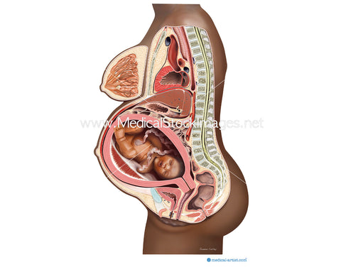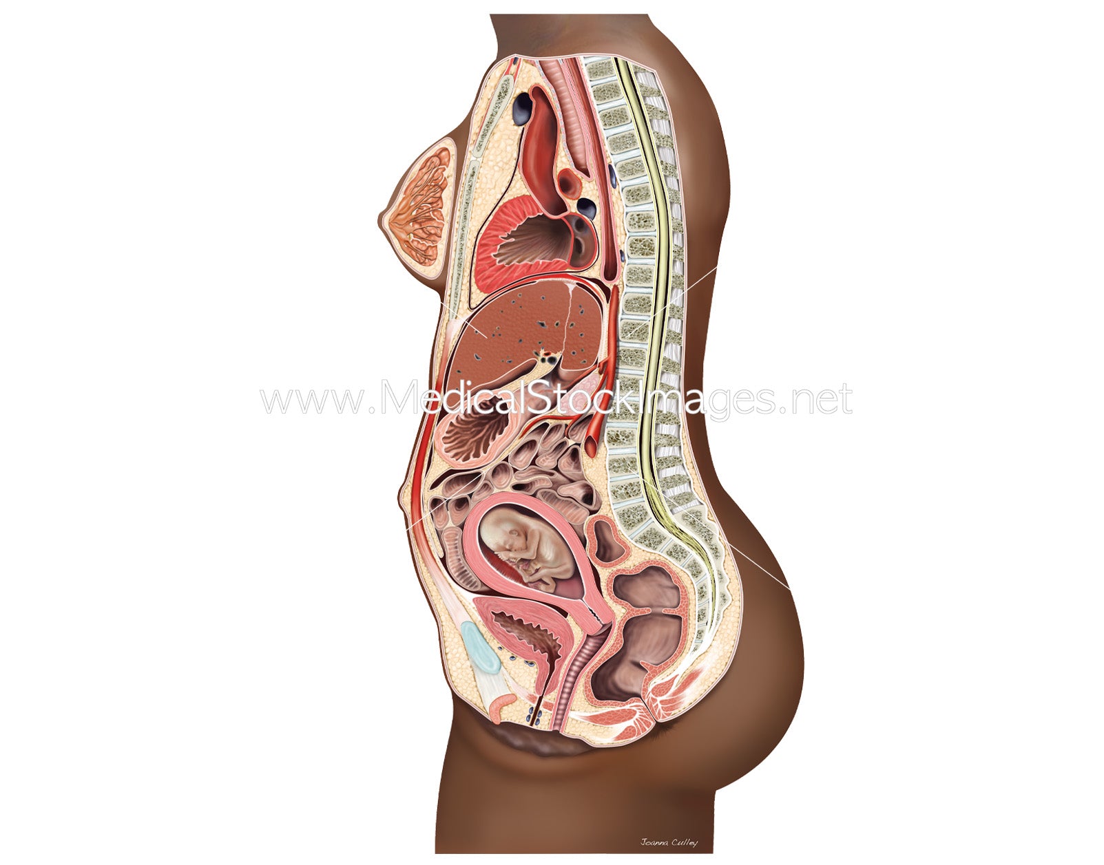
Growing Uterus with Foetal Development at 12 Weeks - Black Baby
Image Description:
The illustration shows a human foetus/fetus within the womb or uterus of a pregnant black woman. The female body has been illustrated in a cross section (cut in half down the middle) to show the foetus/fetus inside the womb as well as the anatomy surrounding the tiny developing baby. The foetus/fetus has not been illustrated in cross section but rather shown as whole and a forming foetus/fetus alongside the placenta at approximately 12 weeks from conception.
The foetus/fetus is shown larger than it may be in actuality, but this was intentional to make the baby more visible to the viewer within the womb. The skin of the foetus/fetus at this time will have a more pale skin tone as it has a thin and translucence skin at this time of development when compared to the mother who is of African heritage.
These illustrations were originally created as part of a commissioned set of foetal development medical illustrations for educational publications for women of colour.
Image File Sizes:
|
Size |
Pixels |
Inches (@300dpi) |
cm (@300dpi) |
|
Small |
325x600px |
1.1"x2" @300dpi |
2.8x5.1cm @300dpi |
|
Medium |
650x1200px |
2x4" @300dpi |
5.5x10.2cm @300dpi |
|
Large |
1301x2400px |
4x8" @300dpi |
11x20.3cm @300dpi |
|
X-Large |
2168x4000px |
7x13" @300dpi |
18.4x33.9cm @300dpi |
|
Maximum |
2426x4475px |
8.1x14.9" @300dpi |
21x38cm @300dpi |
Anatomy Visible in the Medical Illustration Includes:
Image created by:
Infant, baby, mother, child, foetus, fetus, fetal development, foetus development, pregnancy, 12 weeks, mother, birth, foetus anatomy, fetus anatomy, human anatomy
We Also Recommend