Anatomy of the Knee Joint
Image Description:
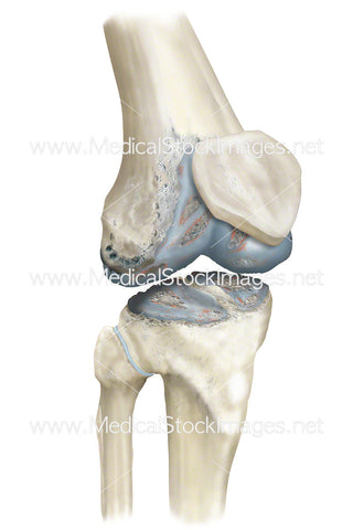
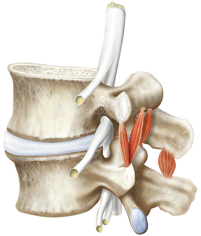
Normal anatomy of the knee joint anterior view. The knee joint is responsible for joining the femur (thigh bone) to the tibia (shin bone) and the femur and the patella (kneecap). Common knee injuries include fractures, dislocations, cruciate ligament injuries and tears to the meniscus and tendons of the knee.
Image File Sizes:
|
Size |
Pixels |
Inches (@300dpi) |
cm (@300dpi) |
|
Small |
377 x 600px |
1.3 x 2” |
3.2 x 5.1cm |
|
Medium |
754 x 1200px |
2.5 x 4” |
6.4 x 10.2cm |
|
Large |
1508 x 2400px |
5 x 8” |
12.8 x 20.3cm |
|
X-Large |
2512 x 4000px |
8.4 x 13.3” |
21.3 x 33.9cm |
|
Maximum |
2734 x 4354px |
9.1 x 14.5” |
23.2 x 36.9cm |
Anatomy Visible in the Medical Illustration Includes:
Anterior cruciate ligament, posterior cruciate ligament, lateral collateral ligament, lateral meniscus, fibula, tibia, medial meniscus, medial collateral ligament, posterior cruciate ligament, femur, thigh bone
Image created by:
We Also Recommend