Trachea and Bronchioles Anatomy
Image Description:
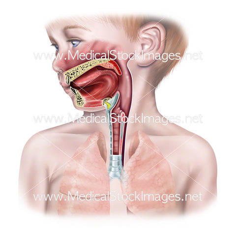
Illustration of the trachea and larynx anterior view showing two principle bronchi dividing into two lobar bronchi for the left lung, and into four lobar bronchi for the right lung. These further divide into smaller bronchi and bronchioles. The trachea is shown with the rings of hyaline cartilage filled by the trachealis muscle, a bundle of smooth muscle and fibroelastic tissue that hold the lumen of the trachea open.
Image File Sizes:
|
Size |
Pixels |
Inches (@300dpi) |
cm (@300dpi) |
|
Small |
600 x 577px |
2.0 x 1.9” |
5.1 x 4.9cm |
|
Medium |
1200 x 1153px |
4.0 x 3.8” |
10.2 x 9.8cm |
|
Large |
2400 x 2305px |
8.0 x 7.7” |
20.3 x 19.5cm |
|
X-Large |
4000 x 3842px |
13.3 x 12.8” |
33.9 x 32.5cm |
|
Maximum |
12992 x 12480px |
43.3 x 41.6” |
110 x 105.7cm |
Anatomy Visible in the Medical Illustration Includes:
Trachea, larynx, thyroid cartilage, median cricothyroid ligament, cricoid cartilage, hyaline cartilage, trachealis muscle, fibroelastic tissue, tracheal cartilages, annular or ring shaped or intercartilaginous ligaments.
Image created by:
We Also Recommend