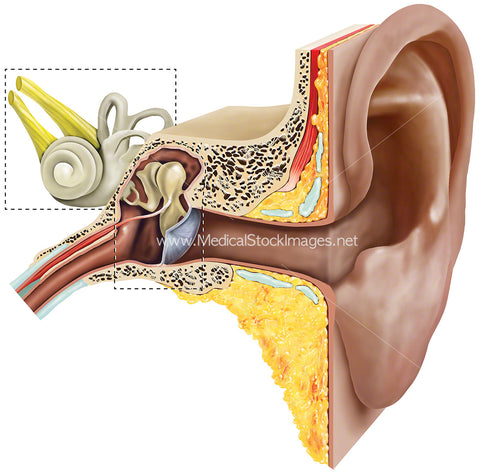
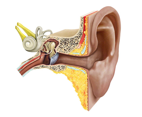
Healthy Ear Cross-Section
Image Description:
This medical illustration shows a healthy human ear in cross-section. The human ear is made up of compartments which is the external, middle, and inner ear. The external ear is called the auricle or pinna and is the visible part of the ear made of ridged cartilage covered by skin. Sound waves are funnelled through the pinna into the external auditory canal to the thin sheet of connective tissue tympanic membrane also known as the eardrum. The mechanical energy of the sound waves causes the eardrum and the attached bones in the middle portion of the ear to vibrate. The cochlea transforms sound into nerve impulses that travel to the brain.
Image File Sizes:
|
Size |
Pixels |
Inches (@300dpi) |
cm (@300dpi) |
|
Small |
600 x 425px |
2.0 x 1.4” |
5.1 x 3.6cm |
|
Medium |
1200 x 849px |
4.0 x 2.8” |
10.2 x 7.2cm |
|
Large |
2400 x 1698px |
8.0 x 5.7” |
20.3 x 14.4cm |
Anatomy Visible in the Medical Illustration Includes:
Outer ear, auricle (ear lobe), external auditory meatus, ear canal, pinna, eardrum, tympanum, tympanic membrane, eustachian tube, middle ear, ossicles, stapes, malleus (hammer), stapes (stirrup), incus (anvil), round window, inner ear, auditory, auditory tube, epitympanic recess, tegmen tympani, limbs of stapes,
Image created by:
We Also Recommend