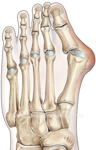
Pencil Drawing of Bony Anatomy of the Foot
Image Description:
Drawn in pencil an illustration of the skeletal foot anatomy with ligaments. Scanned at a very high resolution this drawing is available to print at up to 45cm in height making it ideal for high impact marketing or artwork for medical practices. Illustration is an Anteromedial view.
Image File Sizes:
|
Size |
Pixels |
Inches |
cm |
|
Small |
400x600px |
1.3x2.0” @300dpi |
3.4x5.1cm @300dpi |
|
Medium |
800x1200px |
2.7x4.0” @300dpi |
6.8x10.2cm @300dpi |
|
Large |
1600x2400px |
5.3x8.0” @300dpi |
13.6x20.3cm @300dpi |
|
X-Large |
2667x4000px |
8.9x13.3” @300dpi |
22.6x33.9cm @300dpi |
|
Maximum |
7087x10630px |
23.6x35.4” @300dpi |
60.0x90.0cm @300dpi |
Anatomy Visible in the Medical Illustration Includes:
Fibula, tibia, phalanges: distal, middle and proximal, metatarsal, tarsals: lateral, cuneiform, cuboid, tarsals: medial cuneiform, intermediate cuneiform, navicular, calcaneus, talus, posterior tibiotalar ligament, tibiocalcaneal ligament, anterior tibiotalar ligament, tibionavicular ligament, dorsal cuneonavicular ligament, dorsal intercuneiform ligament, dorsal tarsometatarsals ligament.
Image created by:
We Also Recommend