Illustration of the Skeletal Foot
Image Description:
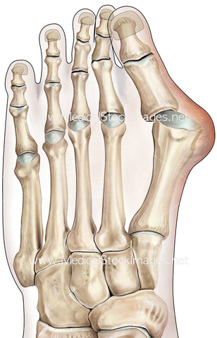
Anatomy of the skeletal foot anterior view. The five toes of the foot contain the phalange bones, the five longer bones connecting to the phalanges are the metatarsal bones. The arches of the feet comprise the tarsals, the three cuneiform bones, the cuboid bone and the navicular bone. The hindfoot comprises the heel (calcaneus bone) and the ankle (talus bone).
Image File Sizes:
|
Size |
Pixels |
Inches |
cm |
|
Small |
404x600px |
1.3x2.0” @300dpi |
3.4x5.1cm @300dpi |
|
Medium |
808x1200px |
2.7x4.0” @300dpi |
6.8x10.2cm @300dpi |
|
Large |
1615x2400px |
5.4x8.0” @300dpi |
13.7x20.3cm @300dpi |
|
X-Large |
2861x4252px |
9.5x14.2” @300dpi |
24.2x36cm @300dpi |
Anatomy Visible in the Medical Illustration Includes:
Phalanges, distal, middle, proximal, metatarsals, tarsals lateral, cuneiform, cuboid, calcaneus, talus, tarsals, navicular, cuneiform, intermediate, medial cuneiform.
Image created by:
We Also Recommend