Full Skeleton with Shoulder, Knee and Foot in Detail
Image Description:
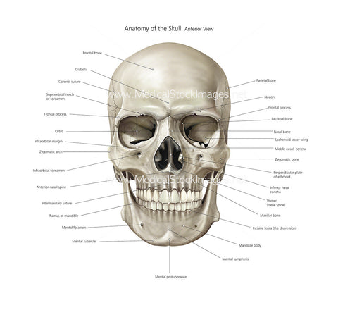
Illustration showing full skeleton and bony anatomy and the shoulder, knee and foot.
Image File Sizes:
|
Size |
Pixels |
Inches (@300dpi) |
cm (@300dpi) |
|
Small |
534 x 600px |
1.8 x 2.0” |
4.5 x 5.1cm |
|
Medium |
1068 x 1200px |
3.6 x 4.0” |
9.1 x 10.2cm |
|
Large |
2136 x 2400px |
7.1 x 8.0” |
18.1 x 20.3cm |
|
X-Large |
3560 x 4000px |
11.9 x 13.3” |
30.1 x 33.9cm |
|
Maximum |
12281 x 13800px |
41.0 x 46.0” |
104.0 x 116.8cm |
Anatomy Visible in the Medical Illustration Includes:
Skull, cranium, mandible, maxilla, sphenoid, zygomatic, clavicle, scapula, sternum, ribs, cervical vertebra, thoracic vertebrae, humerus, radius, ulna, carpals, metacarpals, phalanges, lumbar vertebrae, coccyx, pelvis, pelvic girdle, Ilium, pubis, ischium, femur, tibia, fibula, patella, talus, tarsals, metatarsals, phalanges, frontal bone, nasal bone, parietal bone, temporal bone, lacrimal bone, zygomatic arch, nasal concha, alveolar process, mandible, mental tuberosity, mental protruberance, ramus, nasal spine, volmer, maxilla, ethmoid bone, sphenoid bone, supraorbital foramen, glabella, coronal structure, teeth, thigh bone, distal phalanges, middle phalanges, proximal phalanges, metatarsals, tarsals lateral, cuneiform, cuboid, calcaneus, talus, tarsals, navicular, cuneiform, intermediate, medial cuneiform, tarsal bones, metatarsals, distal phalanx, middle phalanx, proximal phalanx, cuboid, navicular, patella, shoulder capsule, anterior cruciate ligament, posterior cruciate ligament, lateral collateral ligament, lateral meniscus, medial meniscus, medial collateral ligament, posterior cruciate ligament
Image created by:
We Also Recommend