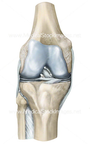
Healthy Knee Lateral Anatomy
Image Description:
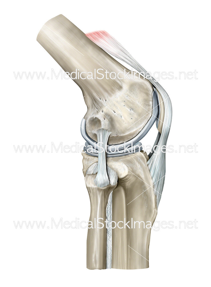
Lateral view of the bony anatomy of the knee including the ligaments and the patella. The ligaments surrounding the knee joint offer stability by limiting movement.
Image File Sizes:
|
Size |
Pixels |
Inches (@300dpi) |
cm (@300dpi) |
|
Small |
442 x 600px |
1.5 x 2.0” |
3.8 x 5.1cm |
|
Medium |
883 x 1200px |
2.9 x 4.0” |
7.5 x 10.2cm |
|
Large |
1765 x 2400px |
5.9 x 8.0” |
15.0 x 20.3cm |
|
X-Large |
2942 x 4000px |
9.8 x 13.3“ |
24.9 x 33.9cm |
|
Maximum |
3361 x 4570px |
11.2 x 15.2” |
28.5 x 38.7cm |
Anatomy Visible in the Medical Illustration Includes:
Femur, quadriceps femoris tendon, patella, articular cartilage, patella ligament, tibia, fibula, interosseous membrane, ligament of fibula head, tibial plateau, lateral meniscus, lateral collateral ligament, lateral epicondyle.
Image created by:
We Also Recommend