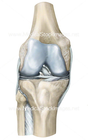
Lateral Healthy Knee - Labelled
Image Description:
Labelled version of the lateral view of the bony anatomy of the knee including the ligaments and the patella. The knee joins the femur to the tibia, these bones, along with the fibula and patella comprise the knee joint.
Image File Sizes:
|
Size |
Pixels |
Inches (@300dpi) |
cm (@300dpi) |
|
Small |
528 x 600px |
1.8 x 2.0” |
4.5 x 5.1cm |
|
Medium |
1056 x 1200px |
3.5 x 4.0” |
8.9 x 10.2cm |
|
Large |
2112 x 2400px |
7.0 x 8.0” |
17.9 x 20.3cm |
|
X-Large |
3099 x 3522px |
10.3 x 11.7” |
26.2 x 29.8cm |
Anatomy Visible in the Medical Illustration Includes:
Femur, quadriceps femoris tendon, patella, articular cartilage, patella ligament, tibia, fibula, interosseous membrane, ligament of fibula head, tibial plateau, lateral meniscus, lateral collateral ligament, lateral epicondyle.
Image created by:
We Also Recommend