Anatomy of a Synovial Joint
Image Description:
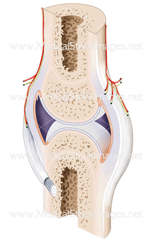
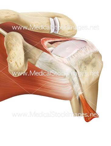
Illustration of a synovial joint, the most common type of joints found in the human body. They consist of cartilage which reduces friction and absorbs shock, synovial fluid which lubricates the joint, synovial membrane which produces synovial fluid, tendons which joins muscles to bones and ligaments which stabilises the joint by joining bone to bone.
Image File Sizes:
|
Size |
Pixels |
Inches (@300dpi) |
cm (@300dpi) |
|
Small |
313 x 600px |
1.0 x 2.0” |
2.7 x 5.1cm |
|
Medium |
843 x 1614px |
2.8 x 5.4” |
7.1 x 13.7cm |
Anatomy Visible in the Medical Illustration Includes:
Synovial joint, bone, blood vessel, nerve, synovial membrane, fibrous capsule, ligament
Image created by:
We Also Recommend

