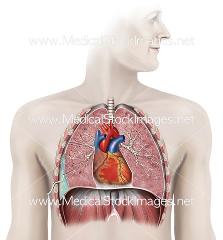
Anatomy of Chest with Drain in place
Image Description:
Illustration showing chest drain in situ with upper chest anatomy. A flexible chest tube is inserted through the chest wall and into the pleural cavity in order to drain air (pneumothorax), blood (haemothorax), fluid (pleural effusion) or pus (empyema) from the chest.
Image File Sizes:
|
Size |
Pixels |
Inches (@300dpi) |
cm (@300dpi) |
|
Small |
559 x 600px |
1.8 x 2.0” |
4.7 x 5.1cm |
|
Medium |
1118 x 1200px |
3.7 x 4.0” |
9.5 x 10.2cm |
|
Large |
2439 x 2618px |
8.1 x 8.7” |
20.7 x 22.2cm |
Anatomy Visible in the Medical Illustration Includes:
Chest drain, lungs, pneumothorax, haemothorax, pleural effusion, empyema, heart, diaphragm
Image created by:
We Also Recommend

