Anatomy of the Inner and Outer Ear
Image Description:
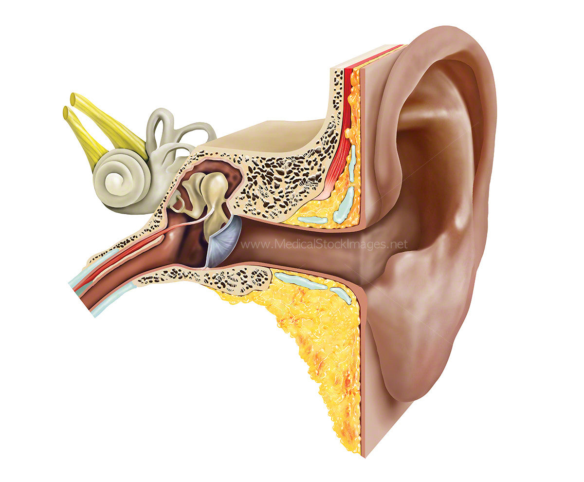
Structures of the ear including the anatomy of the outer, middle ear and inner ear. The ear is composed of three parts all responsible for detecting sound for hearing and to maintain balance.
Image File Sizes:
|
Size |
Pixels |
Inches (@300dpi) |
cm (@300dpi) |
|
Small |
600 x 495px |
2.0 x 1.7” |
5.1 x 4.2cm |
|
Medium |
1200 x 990px |
4.0 x 3.3” |
10.2 x 8.4cm |
|
Large |
2400 x 1981px |
8.0 x 6.6” |
20.3 x 16.8cm |
|
X-Large |
4000 x 3301px |
13.3 x 11.0” |
33.9 x 28.0cm |
|
Maximum |
6096 x 5031px |
20.3 x 16.8” |
51.6 x 42.6cm |
Anatomy Visible in the Medical Illustration Includes:
External ear, auricle, ear canal, outer ear, middle ear, inner ear, ear drum, malleus, semi-circular canals, incus, stapes, vestibular nerve, facial nerve, cochlea, eustachian tube.
Image created by:
We Also Recommend