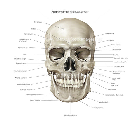
Anatomy of the Skull Anterior View Labelled
Image Description:
An A3 poster sized anatomical illustration of the bones of the skull in anterior view including very detailed labelling of all the visible anatomy. Ideal for use as a teaching aid and available at a high resolution for high quality printing.
Image File Sizes:
|
Size |
Pixels |
Inches |
cm |
|
Small |
600x550px |
2x1.8” @300dpi |
5.1x4.7cm @300dpi |
|
Medium |
1200x1100px |
4x3.7” @300dpi |
10.2x9.3cm @300dpi |
|
Large |
2400x2200px |
8x7.3” @300dpi |
20.3x18.6cm @300dpi |
|
X-Large |
4000x3666px |
13.3x12.2” @300dpi |
33.9x31cm @300dpi |
Anatomy Visible in the Medical Illustration Includes:
Skull, parietal bone, nasion, frontal process, lacrimal bone, nasal bone, spehenoid lesser wing, middle nasal concha, zygomatic bone, perpendicular plate of ethmoid, inferior nasal concha, vomer or nasal spine, maxillar bone, incisive fossa, mandible bone, mental symphysis, mental protuberance, mental tubercle, mental foramen, ramus of mandible, intermaxillary suture, anterior nasal spine, infraorbital foreamen, zygomatic arch, infraorbital margin, orbit, frontal process, supraorbital notch or foreamen, coronal suture, glabella, frontal bone.
Image created by:
We Also Recommend

