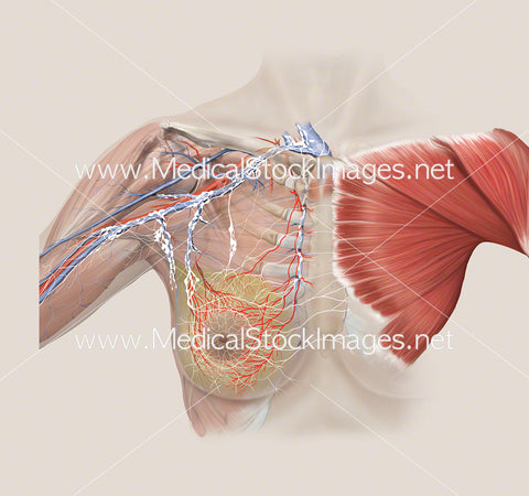
Arterial and Lymphatic Anatomy of Breast with Pectoralis Muscle
Image Description:
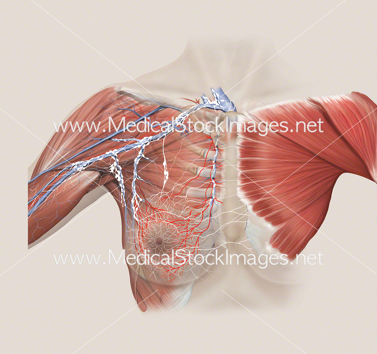
Illustration showing arterial and lymphatic anatomy of the breast with the pectoralis muscle shown on the opposite side.
Image File Sizes:
|
Size |
Pixels |
Inches |
cm |
|
Small |
600x562px |
2.0x1.9” @300dpi |
5.1x4.8cm @300dpi |
|
Medium |
1200x1124px |
4.0x3.7” @300dpi |
10.2x9.5cm @300dpi |
|
Large |
2400x2248px |
8.0x7.5” @300dpi |
20.3x19.0cm @300dpi |
|
X-Large |
3324x3113px |
11.1x10.4" @300dpi |
28.1x26.4cm @300dpi |
Anatomy Visible in the Medical Illustration Includes:
Breast, lateral branches from posterior intercostal arteries, axillary artery, lateral thoracic artery, thoracoacromial artery, deltoid branch, pectoral branch, superior thoracic artery, internal thoracic artery, aortic arch, rib cage, breast tissue, cephalic vein, supraclavicular nodes, interpectoral nodes, Rotter’s nodes, internal mammary nodes, posterior axillary nodes, anterior axillary nodes, lymphatic drainage, Pectoralis muscle
Image created by:
We Also Recommend