Bones of the Foot
Image Description:
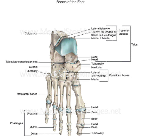
Illustration of a labelled skeleton foot.
Image File Sizes:
|
Size |
Pixels |
Inches |
cm |
|
Small |
600x596px |
2.0x2.0” @300dpi |
5.1x5.1cm @300dpi |
|
Medium |
1200x1192px |
4.0x4.0” @300dpi |
10.2x10.1cm @300dpi |
|
Large |
2400x2385px |
8.0x7.9” @300dpi |
20.3x20.2cm @300dpi |
|
X-Large |
3959x3934px |
13.2x13.1” @300dpi |
33.5x33.3cm @300dpi |
Anatomy Visible in the Medical Illustration Includes:
Calcaneus, talocalcaneonavicular joint, cuboid, tuberosity, metatarsal bones, phalanges, proximal, middle, distal, groove for tendon of flexor hallucis longus, lateral tubercle, medial tubercle, posterior process, talus, trochlea, neck, head, tuberosity, navicular, cuneiform bones, base, body.
Image created by:
We Also Recommend

