Extreme Case of Lumbar 4 and Lumbar 5 Bone Degeneration - White Background
Image Description:
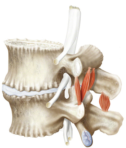
The illustrations depicts an extreme case of bone degeneration located on the L4 and L5 area of the lumbar spine on a white background.
Further anatomy detail includes the interspinal muscle. There are paired muscles located between the spinous processes of adjacent vertebrae; subdivided into cervical, thoracic, and lumbar muscles. The interspinal muscles are short bands of muscle fibres including the interspinales cervicis, interspinales thoracis, and interspinales lumborum muscles.
Protected by the vertebrae, the spinal cord splits out between the vertebrae L1 and L2. The nervous tissue that extends below this point are individual strands that collectively form the cauda equina. In between each lumbar vertebra a nerve root exits, coming together again to form the largest single nerve in the human body, the sciatic nerve. Any disorders of the back may be of a result of this sciatic nerve having become trapped through spinal disc herniation that can affect the nerve root. Pain manifests itself as lower back pain, that radiates along the sciatic nerve through the back of the leg and down to the foot.
Image File Sizes:
|
Size |
Pixels |
Inches |
cm |
|
Small |
512x600px |
1.707x2” @300dpi |
4.33x5.08cm @300dpi |
|
Medium |
1023x1200px |
3.41x4” @300dpi |
8.66x10.16cm @300dpi |
|
Large |
2046x2400px |
6.82x8” @300dpi |
17.32x20.32cm @300dpi |
|
X-Large |
3069x3600px |
10.23x12” @300dpi |
25.98x30.48cm @300dpi |
Anatomy Visible in the Medical Illustration Includes:
Lumbar, vertebra, vertebrae, L4, L5, spine, degeneration, bone, interspinales, muscles,
Image created by:
We Also Recommend

