Female Urinary Anatomy
Image Description:
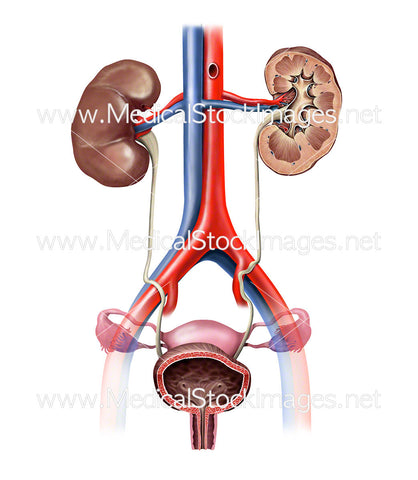
Female urinary anatomy anterior view created in digital colour. Includes surface anatomy of one kidney and cross-section view of other kidney. The kidneys filter waste products and water to form urine, which moves to the bladder via tiny tubes called ureters. Urine leaves the bladder through the urethra.
Image File Sizes:
|
Size |
Pixels |
Inches (@300dpi) |
cm (@300dpi) |
|
Small |
522 x 600px |
1.7 x 2.0” |
4.4 x 5.1cm |
|
Medium |
1044 x 1200px |
3.5 x 4.0” |
8.8 x 10.2cm |
|
Large |
1995 x 2294px |
6.7 x 7.6” |
16.9 x 19.4cm |
Anatomy Visible in the Medical Illustration Includes:
Bladder, kidney, kidney in cross section, ureter, right kidney surface anatomy, uterus, fallopian tubes, urethra, common iliac artery, common iliac vein, renal artery, renal vein.
Image created by:
We Also Recommend

