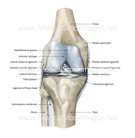
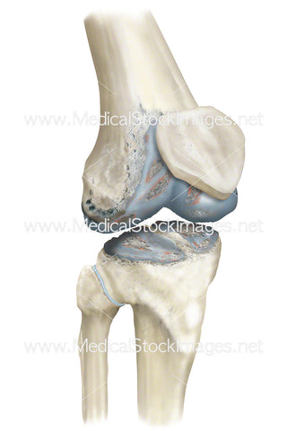
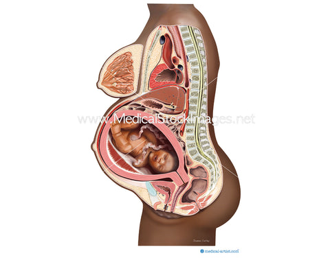
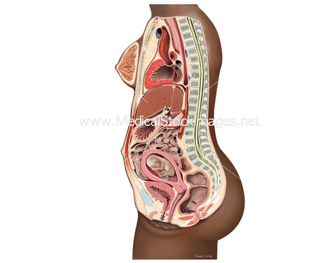
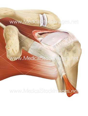
Healthy Knee Anterior View Labelled
Image Description:
Anatomy of the knee anterior view and its ligaments. The two important ligaments of the knee – the anterior cruciate ligament (ACL) and posterior cruciate ligament (PCL) – are crucial to the stability of the knee.
Image File Sizes:
|
Size |
Pixels |
Inches (@300dpi) |
cm (@300dpi) |
|
Small |
588 x 600px |
2.0 x 2.0” |
5.0 x 5.1cm |
|
Medium |
1175 x 1200px |
3.9 x 4.0” |
9.9 x 10.2cm |
|
Large |
2349 x 2400px |
7.8 x 8.0” |
19.9 x 20.3cm |
|
X-Large |
3547 x 3624px |
11.8 x 12.1” |
30 x 30.7cm |
Anatomy Visible in the Medical Illustration Includes:
Femur, medical epicondyle, medial collateral ligament, posterior cruciate ligament, transverse ligament, tibia, fibula, interosseous membrane, ligament of fibula head, tibial plateau, lateral meniscus, anterior cruciate ligament, lateral cruciate ligament, articular cartilage, patellofemoral groove.
Image created by:
We Also Recommend

