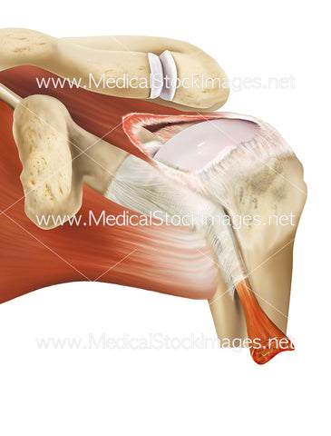
Labelled Anatomy of the Muscles of the Back
Image Description:
Labelled illustration of the superficial muscles of the back. The three major groups comprising the back muscles are the superficial, intermediate and deep muscles. The superficial muscles are most commonly associated with the movements of the shoulder, attaching to the clavicle, scapula and humerus.
Image File Sizes:
|
Size |
Pixels |
Inches (@300dpi) |
cm (@300dpi) |
|
Small |
585 x 600px |
2.0 x 2.0” |
5.0 x 5.1cm |
|
Medium |
1170 x 1200px |
3.9 x 4.0” |
9.9 x 10.1cm |
|
Large |
2341 x 2400px |
7.8 x 8.0” |
19.8 x 20.3cm |
|
X-Large |
4571 x 4687px |
15.2 x 15.6” |
38.7 x 39.7cm |
Anatomy Visible in the Medical Illustration Includes:
Trapezius, deltoid, triceps brachii, teres major, infranspinatus, rhomboid major, latissimus dorsi, thoracolumbar fascia, external oblique, internal oblique, gluteus.
Image created by:
We Also Recommend


