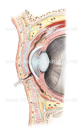
Orbital Cavity of the Eye (Sagittal View)
Image Description:
The orbit is the bony cavity in the skull where the eye is situated. It is also known as the eye socket and comprises of seven conjoined bones which protect and support the function of the eye.
Image File Sizes:
|
Size |
Pixels |
Inches (@300dpi) |
cm (@300dpi) |
|
Small |
367 x 600px |
1.2 x 2.0” |
3.1 x 5.1cm |
|
Medium |
733 x 1200px |
2.4 x 4.0” |
6.2 x 10.2cm |
|
Large |
1466 x 2400px |
4.9 x 8.0” |
12.4 x 20.3cm |
|
X-Large |
2403 x 3933px |
8.0 x 13.1” |
20.4 x 33.3cm |
Anatomy Visible in the Medical Illustration Includes:
Orbital septum, orbicularis oculi, upper eyelid, ciliary, sebaceous glands, lower eyelid, orbital roof, periorbita, levator palpebrae superioris, superior rectus, superior conjunctival fornix, superior tarsal muscle, superior tarsus, tarsal glands, lens, cornea, iris, ciliary body, inferior tarsus, retina, sclera, inferior tarsal muscle, obicularis oculi, infraorbital nerve
Image created by:
We Also Recommend

