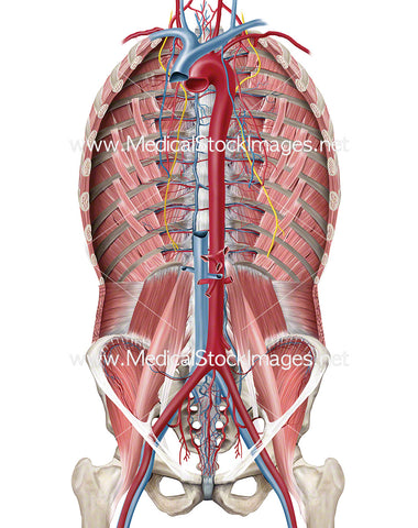
Arteries and Veins of the Ventral Cavity including the Phrenicus Nerve
Image Description:
Muscle of the posterior abdominal wall without the diaphragm. Vessels and phrenic nerve (nervus phrenicus) present. The phrenic nerve originates in the neck and is responsible for innervating the diaphragm.
Image File Sizes:
|
Size |
Pixels |
Inches |
cm |
|
Small |
476x600px |
1.6x2.0” @300dpi |
4.0x5.1cm @300dpi |
|
Medium |
951x1200px |
3.2x4.0” @300dpi |
8.1x10.2cm @300dpi |
|
Large |
1901x2400px |
6.3x8.0” @300dpi |
16.1x20.3cm @300dpi |
|
X-Large |
3168x4000px |
10.6x13.3” @300dpi |
26.8x33.9cm @300dpi |
|
Maximum |
6916x8731px |
23.1x29.1” @300dpi |
58.6x73.9cm @300dpi |
Anatomy Visible in the Medical Illustration Includes:
Internal intercostal muscles, transverse abdominis, psoas major, iliacus, iliopsoas, psoas minor, quadratus lumborum, pelvis, rib cage in cross section, phrenicus nerve.
Image created by:
We Also Recommend


