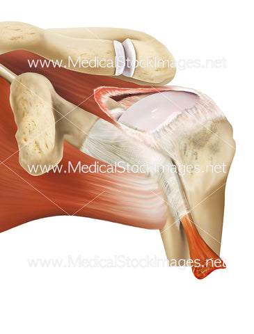
Skeletal and Muscular Anatomy of the Shoulder
Image Description:
Anterior view of the skeleton of the right upper limb and anterior view of the muscles of the right upper limb. The bones of the shoulder consist of the scapula, the humerus and the clavical. These three bones together with the ball and socket joint allow the shoulder a large range of motion such as adduction, abduction, flexion, extension, internal rotation, external rotation, and 360° circumduction in the sagittal plane. The elbow joint is a hinge joint formed between the distal end of the humerus and the proximal ends of the ulna and radius and being a hinge joint, the only movements allowed by the elbow are flexion and extension.
Image File Sizes:
|
Size |
Pixels |
Inches (@300dpi) |
cm (@300dpi) |
|
Small |
600 x 547px |
2.0 x 1.8” |
5.1 x 4.6cm |
|
Medium |
1200 x 1093px |
4.0 x 3.6” |
10.2 x 9.3cm |
|
Large |
2400 x 2186px |
8.0 x 7.3” |
20.3 x 18.5cm |
|
X-Large |
3294 x 3000px |
11.0 x 10.0” |
27.9 x 25.4cm |
Anatomy Visible in the Medical Illustration Includes:
Clavicle, scapula, acromioclavicular joint, elbow joint: humerocradial joint, humeroulnar joint, proximal radioulnar joint, humerus, radius, ulna, Supraspinatus, serratus anterior, subscapularis, teres major, pronator teres, common head of flexors, brachialis, bicipital aponeurosis, biceps brachii, tendon of insertion, biceps brachii long head, biceps brachii short head, pectoralis major, coracobrachialis, pectoral minor.
Image created by:
We Also Recommend


