Skeletal and Muscular Anatomy of the Shoulder
Image Description:
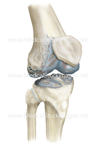
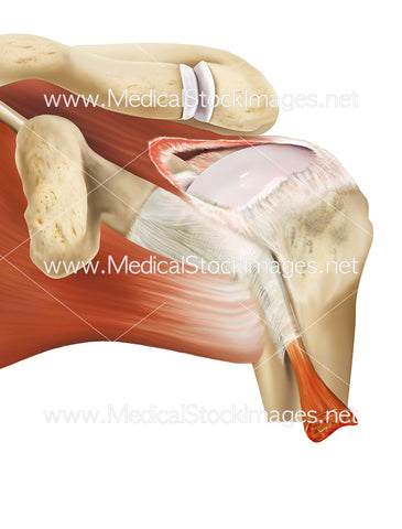
Illustrations showing the skeletal and muscular anatomy of the shoulder.
Image File Sizes:
|
Size |
Pixels |
Inches (@300dpi) |
cm (@300dpi) |
|
Small |
600 x 499px |
2.0 x 1.7” |
5.1 x 4.2cm |
|
Medium |
1200 x 998px |
4.0 x 3.3” |
10.2 x 8.5cm |
|
Large |
2400 x 1996px |
8.0 x 6.7” |
20.3 x 16.9cm |
|
X-Large |
4000 x 3326px |
13.3 x 11.1” |
33.9 x 28.2cm |
|
Maximum |
5985 x 4977px |
20.0 x 16.6” |
50.7 x 42.1cm |
Anatomy Visible in the Medical Illustration Includes:
Supraspinatus, clavicle, serratus anterior, subscapularis, teres major, pronator teres, common head of flexors, brachialis, bicipital aponeurosis, biceps brachii, tendon of insertion, biceps brachii long head, biceps brachii short head, pectoralis major, coracobrachialis, pectoral minor, acromion, coracoid process, humerus, radius, ulna, clavicle, glenoid, scapula, humeroradial, humeroulnar, radioulnar, elbow
Image created by:
We Also Recommend


