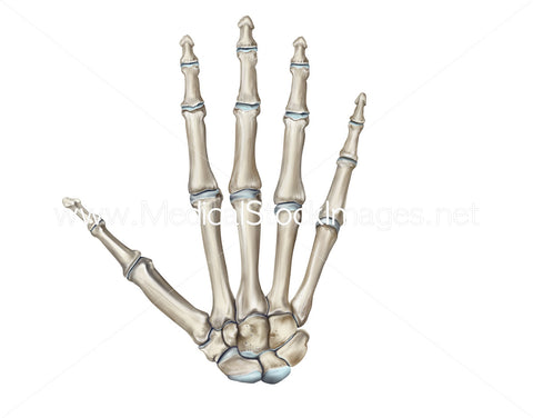
Skeleton Hand-Bones of the Hand Dorsal View
Image Description:
An anatomically precise illustration of the palmer view of the bones of the hand.
Image File Sizes:
|
Size |
Pixels |
Inches (@300dpi) |
cm (@300dpi) |
|
Small |
496 x 600px |
1.7 x 2.0” |
4.2 x 5.1cm |
|
Medium |
991 x 1200px |
3.3 x 4.0” |
8.4 x 10.2cm |
|
Large |
1982 x 2400px |
6.6 x 8.0” |
16.8 x 20.3cm |
|
X-Large |
3303 x 4000px |
11.0 x 13.3” |
28.0 x 33.9cm |
|
Maximum |
5058 x 6125px |
16.9 x 20.4” |
42.8 x 51.9cm |
Anatomy Visible in the Medical Illustration Includes:
Phalanges, metacarpal bones, carpal bones, second distal phalanx, second middle phalanx, second proximal phalanx, first distal phalanx, second proximal phalanx, sesamoid bones, first metacarpal, shaft of metacarpal, head of metacarpal, base of phalanx, shaft of phalanx, head of phalanx, tuberosity of distal phalanx, hamate, scaphoid, lunate, triquetrum, pisiform, capitate, hook of hamate, tubercle of trapezium, trapezium, trapezoid.
Image created by:
We Also Recommend

