Spinal Cord in Posterolateral View
Image Description:
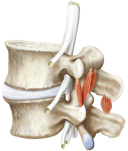
Illustration showing the spine and spinal cord in posteriolateral view. The spinal cord runs from the base of the brain to the lower part of the thoracic spine. There are 8 cervical nerves, 12 thoracic nerves, 5 lumbar nerves, 5 sacral nerves and 1 coccygeal nerve. The spinal cord, contains neurons that carry sensory information from the body to the central nervous system, allowing for processing of the information received and also carry information from the head to the central nervous system, allowing motor functions to be performed. Any damage to the spinal cord can result in loss of sensory and/or motor function below the level of injury.
Image File Sizes:
|
Size |
Pixels |
Inches |
cm |
|
Small |
541x600px |
1.8x2.0” @300dpi |
4.6x5.1cm @300dpi |
|
Medium |
1082x1200px |
3.6x4.0” @300dpi |
9.2x10.2cm @300dpi |
|
Large |
2165x2400px |
7.2x8.0” @300dpi |
18.3x20.3cm @300dpi |
|
X-Large |
3608x4000px |
12.0x13.3” @300dpi |
30.6x33.9cm @300dpi |
|
Maximum |
4382x4858px |
14.6x16.2” @300dpi |
37.1x41.1cm @300dpi |
Anatomy Visible in the Medical Illustration Includes:
Spine, spinal cord, spinal nerve, exiting nerve roots, nerve roots, spinal ganglion, pia mater, dura mater, azygos vein, arachnoid mater, thoracic duct, rhomboid major m., trapezius muscle, aorta, thoracic 6, thoracic 7.
Image created by:
We Also Recommend