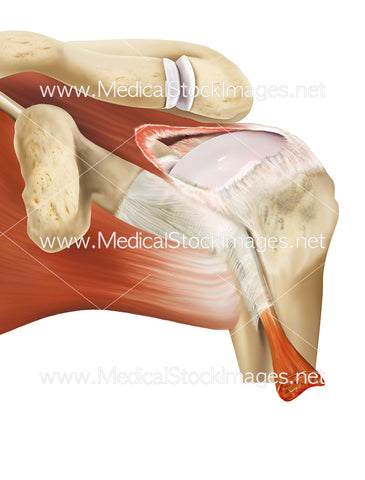
Superficial Muscles of the Posterior of the Head and Neck
Image Description:
Illustration showing the posterior view of head and neck with superficial muscles.
Image File Sizes:
|
Size |
Pixels |
Inches (@300dpi) |
cm (@300dpi) |
|
Small |
600 x 474px |
2.0 x 1.6” |
5.1 x 4.1cm |
|
Medium |
1200 x 947px |
4.0 x 3.2” |
10.2 x 8.0cm |
|
Large |
2400 x 1894px |
8.0 x 6.3” |
20.3 x 16.0cm |
|
X-Large |
3167 x 2499px |
10.6 x 8.3” |
26.8 x 21.2cm |
Anatomy Visible in the Medical Illustration Includes:
Head, neck, muscles, sternocleidomastoid, thoracolumbar fascia, deep layer of nuchal faschia, rhomboid minor, levator scapulae, clavicle, acromion, supraspinatus, trapezius, descending, transverse, scapular spine.
Image created by:
We Also Recommend


